
by Terri Malloy,Mayo Clinic
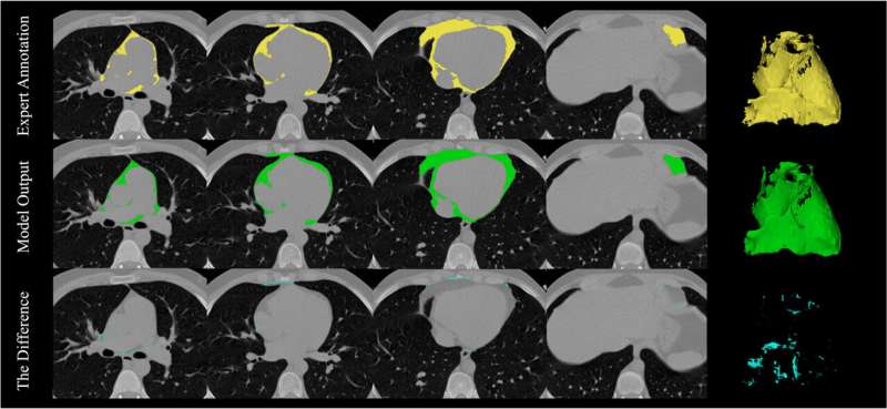
Pericardial adipose tissue (PAT) segmentation: A visual comparison of the ground truth segmentations of the PAT (upper row), model’s predictions (middle row) and the subtraction of two masks. From left to the right, columns represent 2D axial slices from upper to lower heart levels and the rightmost column corresponds to the 3D representation of the masked region. Credit:American Journal of Preventive Cardiology(2026). DOI: 10.1016/j.ajpc.2026.101549
Mayo Clinic research has identified a powerful new way to improve the prediction of a patient's long-term cardiovascular disease risk by enhancing a routinely performed imaging test with artificial intelligence (AI). Heart disease develops over time and remains the leading cause of death worldwide, so identifying risk early is critical to preventing heart attack, stroke, and other serious outcomes.
The study highlights the growing role of AI in helping experts uncover new insights from existing medical data. Findings were presented at the2026 American College of Cardiology Scientific Sessionwith simultaneouspublicationin theAmerican Journal of Preventive Cardiology.
The study followed nearly 12,000 adults for approximately 16 years. Investigators applied AI to participants' standardcoronary artery calciumscans to measure fat surrounding the heart. They compared the predictive value of this measurement with and in combination with two standard risk assessment approaches: theAmerican Heart Association PREVENT equation, which incorporates traditional factors such as age, sex, blood pressure, cholesterol, diabetes, and other variables; and the coronary artery calcium score, which measures calcified plaque in coronary arteries.
The findings show that the volume of heart fat could be used independently to predict cardiovascular events. It significantly improved the overall accuracy of long-term risk prediction when combined with the coronary artery calcium score and the PREVENT equation, especially among patients in low-risk categories.
"Pericardial fathas been recognized as a marker of cardiovascular risk, but this study shows how we can now measure it automatically and use it to meaningfully improve risk prediction, especially in patients at borderline or intermediate risk where clinical decisions are often less clear," says Zahra Esmaeili, first author and researcher in the Department of Cardiovascular Medicine at Mayo Clinic. "This opens the door to more personalized prevention strategies."
Key findings:
Coronary artery calcium scoring is widely used to assess cardiovascular risk. This study shows that additional information can be extracted from the same scan without extra testing or cost.
"Because this measurement comes from imaging that many patients are already receiving, it represents apractical and scalableway to enhance cardiovascular risk assessment," says senior author Francisco Lopez-Jimenez, M.D., a preventive cardiologist and co-director of the AI in Cardiology program at Mayo Clinic. "It could help clinicians intervene earlier and more effectively."
Researchers note that further studies will help determine how best to incorporate coronary fat measurement into routine clinical care and whether it can guide treatment decisions.
More information Zahra Esmaeili et al, Deep learning-derived pericardial adipose tissue by electrocardiogram-gated cardiac computed tomography predicts cardiovascular events beyond coronary calcium score, American Journal of Preventive Cardiology (2026). DOI: 10.1016/j.ajpc.2026.101549


Post comments