

Introduction
Breast cancer continues to be one of the leading causes of mortality among women worldwide, and the value of early detection in improving survival rates cannot be overstated. Mammography remains the most widely adopted screening tool and has saved millions of lives through early diagnosis. Yet, it is not without significant limitations. Breast compression causes discomfort for many women, repeated exposure to ionizing radiation raises concerns about cumulative risks, and diagnostic accuracy is reduced in women with dense breast tissue. Moreover, mammography requires costly infrastructure and trained personnel, making it less accessible in low- and middle-income regions. These limitations have spurred the search for safe, affordable, and widely deployable alternatives that can complement existing methods.
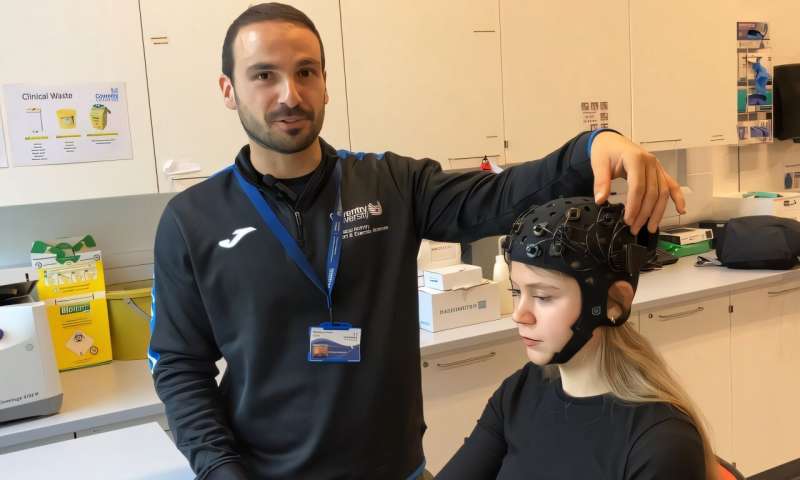
One such technology is infrared thermography (IRT). Once dismissed for its interpretive limitations, IRT is now experiencing a revival. The method is entirely non-invasive, radiation-free, and capable of detecting subtle physiological changes associated with malignancies. Recent advances in artificial intelligence, particularly the development of multi-convolutional neural networks (multi-CNNs), have transformed thermography into a powerful diagnostic tool capable of distinguishing malignant from benign thermal patterns with unprecedented accuracy. Together, IRT and AI represent an emerging frontier in breast cancer screening that could reshape how early detection is practiced worldwide.
Infrared Thermography in Breast Cancer Detection
Infrared thermography measures heat emitted from the surface of the skin, capturing thermal variations that reflect underlying physiological activity. Malignant tumors are characterized by increased vascularization and higher metabolic activity, which generate localized regions of elevated temperature. These “hotspots” can be visualized on thermal images, offering a window into processes not visible through conventional imaging.
Compared with mammography, IRT offers multiple advantages. It does not expose patients to radiation, it is painless and non-contact, and it can be repeated as often as needed without risk. Its portability and relatively low cost also make it suitable for use in resource-limited regions where mammography infrastructure is unavailable. Younger women, who are typically excluded from routine mammography due to radiation exposure concerns, may also benefit from IRT as a safer monitoring option.
Nevertheless, IRT historically faced challenges that hindered its adoption. Early interpretation relied heavily on human observation, leading to high false-positive rates because benign conditions such as infections or hormonal fluctuations could mimic malignant signatures. This interpretive weakness restricted its clinical utility for decades. The emergence of artificial intelligence, however, has revolutionized thermography by providing the ability to analyze complex thermal patterns with far greater precision.
The Role of Multi-Convolutional Neural Networks
Convolutional neural networks have become a cornerstone of modern medical imaging because of their ability to automatically extract features and recognize patterns beyond human visual capacity. In breast cancer thermography, multi-CNN architectures are particularly effective. Instead of relying on a single neural network to process an image, multiple CNN streams are employed to analyze different aspects of the thermogram. One stream might focus on overall thermal distribution, another on localized anomalies, and a third on segmented regions of interest. By fusing these outputs, multi-CNNs achieve a more comprehensive and reliable classification.
This multi-stream approach directly addresses thermography’s historical limitations. By analyzing both global and local features, the networks can differentiate between malignant and benign anomalies with remarkable accuracy. Recent studies have demonstrated that ensembles of advanced CNN architectures such as ResNet, DenseNet, and EfficientNet, when applied to thermal imaging datasets, achieve diagnostic accuracies exceeding 96 percent, with improvements in both sensitivity and specificity. These results are especially striking given that conventional thermography was once criticized for its unreliability.
Beyond accuracy, multi-CNNs also provide robustness against noise and variability in thermal images, further improving their clinical potential. By integrating clinical metadata such as patient age, family history, or hormonal status, these models can contextualize thermal signatures and improve decision-making. The combination of thermography and AI is thus not only technologically innovative but also clinically meaningful, offering a diagnostic pathway that is both patient-centered and adaptable to diverse contexts.
From Thermogram to Diagnosis
The application of IRT with multi-CNNs follows a structured process. Thermal images of the breast are first acquired under standardized environmental conditions to ensure reproducibility and accuracy. These raw images are then preprocessed through calibration, noise reduction, and segmentation, ensuring that regions of interest are accurately isolated for analysis. Once prepared, the images are fed into multiple CNN streams, each designed to detect specific types of thermal features, from broad distribution patterns to localized hotspots.
The networks extract features automatically, and their outputs are integrated to produce a probabilistic classification of the breast tissue, distinguishing benign lesions from potential malignancies. In some cases, additional clinical data are incorporated to contextualize the findings. The final stage involves synthesizing the results into a diagnostic report, highlighting suspicious areas and providing confidence scores that support clinical decision-making.
This end-to-end pipeline is not merely a technical process but a clinically viable workflow. It allows for the translation of raw thermal data into actionable insights, offering clinicians a complementary tool alongside mammography. Early trials and pilot studies have shown that this approach achieves high sensitivity and specificity, with some models reporting accuracies close to 99 percent.
Clinical Promise and Remaining Challenges
The promise of IRT and multi-CNNs lies in their ability to democratize access to breast cancer screening. Portable, non-invasive, and affordable, this approach is particularly well suited for low-resource settings where mammography infrastructure is limited or unavailable. For younger women and those requiring frequent monitoring, thermography offers a safe alternative without radiation risks. In clinical environments, it has the potential to function as a complementary modality, improving diagnostic confidence and reducing reliance on a single imaging method.
Yet challenges remain before this technology can be fully integrated into routine practice. One of the most pressing needs is the establishment of standardized imaging protocols to ensure consistency across institutions. In addition, AI models require large, diverse, and well-annotated datasets to achieve generalizable accuracy across populations. Without such datasets, there is a risk of bias in diagnostic outcomes, particularly across different ethnic groups and geographic regions. Clinical validation through large-scale trials, regulatory approval, and training programs for healthcare professionals will also be necessary to translate this promising technology into practice.
Conclusion
Breast cancer screening stands at a pivotal moment. While mammography remains indispensable, its limitations highlight the importance of pursuing complementary methods that are safe, accessible, and accurate. Infrared thermography, once sidelined due to interpretive challenges, has been revitalized by advances in artificial intelligence. Multi-convolutional neural networks now allow thermography to achieve high diagnostic accuracy, transforming it into a clinically viable tool for early detection.
The integration of IRT with multi-CNNs represents more than a technological advancement; it signals a shift toward more inclusive and patient-centered screening strategies. By making early detection safer, more comfortable, and more accessible across diverse populations, AI-enhanced thermography could help bridge existing inequities in cancer care. Its role is not to replace mammography, but to complement it, expanding the range of diagnostic tools available to clinicians and patients. As research continues, this synergistic approach has the potential to reshape the future of breast cancer screening, offering hope for earlier detection and better outcomes for women worldwide.
Reference
1.Rakhunde MB, Gotarkar S, Choudhari SG. Thermography as a Breast Cancer Screening Technique: A Review Article. Cureus. 2022 Nov 8;14(11):e31251. doi: 10.7759/cureus.31251. PMID: 36505165; PMCID: PMC9731505.
2.Attallah O. Harnessing infrared thermography and multi-convolutional neural networks for early breast cancer detection. Sci Rep. 2025 Jul 28;15(1):27464. doi: 10.1038/s41598-025-09330-2. PMID: 40721416; PMCID: PMC12304260.
3.Ng EY, Kee EC. Advanced integrated technique in breast cancer thermography. J Med Eng Technol. 2008 Mar-Apr;32(2):103-14. doi: 10.1080/03091900600562040. PMID: 17852648.
4.Asok Bandyopadhyay, Himanka S. Mondal, Bivas Dam, Dipak C. Patranabis, Barnali Pal. Innovative infrared imaging approach for breast cancer screening: Integrating rotational thermography and machine learning analysis. Artificial Intelligence in Health 2024, 1(3), 64–79. https://doi.org/10.36922/aih.3312
5.Khan AQ, Touseeq M, Rehman S, Tahir M, Ashfaq M, Jaffar E and Abbasi SF (2025) Advances in breast cancer diagnosis: a comprehensive review of imaging, biosensors, and emerging wearable technologies. Front. Oncol. 15:1587517. doi: 10.3389/fonc.2025.1587517`
6.Li M, Jiang Y, Zhang Y, Zhu H. Medical image analysis using deep learning algorithms. Front Public Health. 2023 Nov 7;11:1273253. doi: 10.3389/fpubh.2023.1273253. PMID: 38026291; PMCID: PMC10662291.
7.Chao Chen, Nor Ashidi Mat Isa, Xin Liu, A review of convolutional neural network based methods for medical image classification, Computers in Biology and Medicine, Volume 185,
8.Amreen Batool, Yung-Cheol Byun, A lightweight multi-path convolutional neural network architecture using optimal features selection for multiclass classification of brain tumor using magnetic resonance images, Results in Engineering,
9.Ahmed KS, Sherif FF, Abdallah MS, Cho YI, ElMetwally SM. An Innovative Thermal Imaging Prototype for Precise Breast Cancer Detection: Integrating Compression Techniques and Classification Methods. Bioengineering (Basel). 2024 Jul 29;11(8):764. doi: 10.3390/bioengineering11080764. PMID: 39199722; PMCID: PMC11352007.
10.Alzahrani, R.M., Sikkandar, M.Y., Begum, S.S. et al. Early breast cancer detection via infrared thermography using a CNN enhanced with particle swarm optimization. Sci Rep15, 25290 (2025). https://doi.org/10.1038/s41598-025-11218-0
11.Wang X, Chou K, Zhang G, Zuo Z, Zhang T, Zhou Y, Mao F, Lin Y, Shen S, Zhang X, Wang X, Zhong Y, Qin X, Guo H, Wang X, Xiao Y, Yi Q, Yan C, Liu J, Li D, Liu W, Liu M, Ma X, Tao J, Sun Q, Zhai J, Huang L. Breast cancer pre-clinical screening using infrared thermography and artificial intelligence: a prospective, multicentre, diagnostic accuracy cohort study. Int J Surg. 2023 Oct 1;109(10):3021-3031. doi: 10.1097/JS9.0000000000000594. PMID: 37678284; PMCID: PMC10583949.


Post comments