
bySPIE
Label-free multiphoton microscopy and deep learning can be used in combination to classify pancreatic neuroendocrine neoplasms with high accuracy—a significant step toward automated digital pathology for such tumors. Credit: N. Daigle (University of Arizona).
Pancreatic neuroendocrine neoplasms (PNENs) are a rare form of cancer that affects hormone-producing cells in the pancreas. Although uncommon, their incidence has been rising steadily over the past few decades.
Treatment options include chemotherapy and targeted therapies, but surgery remains the only chance for a cure. However, surgical decisions often depend on pathology results that can take hours or even days, delaying treatment and increasing the risk of incomplete tumor removal.
Researchers at the University of Arizona have developed a new imaging method that could help surgeons identifycancerous tissuemore quickly and accurately.
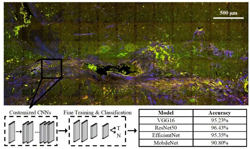
The technique, calledmultiphoton microscopy(MPM), uses a form of light-based imaging that can highlight natural fluorescent molecules in tissue. Unlike traditional microscopy, MPM causes less damage to samples and provides clearer images, making it a promising tool for real-time analysis during surgery.
AsreportedinBiophotonics Discovery, the research team used MPM to scan pancreatic tissue samples for naturally occurring fluorescent markers like collagen, NADH, FAD, lipofuscins, and porphyrins.
These markers help distinguish between healthy and cancerous tissue. To interpret the images, the researchers applied both machine learning (ML) anddeep learning techniques. One ML algorithm and fourconvolutional neural networks(CNNs) were trained to classify the tissue types.
The results were encouraging. The ML algorithm achieved an accuracy of 80.6% in identifying cancerous tissue, while the CNNs performed even better, with accuracies ranging from 90.8% to 96.4%. These high scores are especially notable because the samples came from multiple biorepositories, suggesting that the method is robust across different sources.
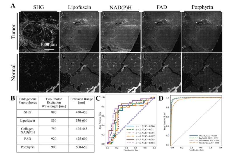
(A) Pancreatic neuroendocrine neoplasms (PNENs) and normal pancreatic tissue samples were imaged (B) at five different wavelengths to capture changes in tissue autofluorescence as a result of cancer. Results showed that (C) machine learning was able to classify the tissue types with good accuracy as the number of features given to the algorithm increased up to six, but (D) convolutional neural networks (CNNs) had much higher accuracy. Credit: N. Daigle et al., DOI: 10.1117/1.BIOS.2.4.045001.
While the CNNs outperformed the ML algorithm, the latter offered more transparency. By analyzing which features influenced the ML model's decisions, the researchers found that collagen content and image characteristics like contrast and correlation were key indicators of cancer.
This insight could help refine future models and improve understanding of PNEN tissue structure.
The study also showed that MPM imaging is faster than traditional histology, though the researchers believe further improvements could make it even quicker. Next, they plan to test the technique on fresh tissue samples during surgery and explore whether it can help determine the grade and type of PNENs—information that could guide treatment decisions more precisely.
This research points to a future wherecancer diagnosisand surgical planning could happen in near real-time, potentially reducing the need for repeat surgeries and improving outcomes for patients withpancreatic cancer.
More information: Noelle Daigle et al, Investigating machine learning algorithms to classify label-free images of pancreatic neuroendocrine neoplasms, Biophotonics Discovery (2025). DOI: 10.1117/1.bios.2.4.045001
Provided by SPIE


Post comments