
A procedure to look at blood flow inside the body
By Brandon Peters, MD
Updated on October 03, 2023
Medically reviewed by Richard N. Fogoros, MD
Angiography is a medical procedure used to visualize blood flow in the body using imaging techniques, like an X-ray. During an angiogram, contrast substances are injected into the blood to better show how it flows. This can help with diagnosing and treating various medical conditions, especially those that impact the heart and brain.
Angiography is not considered a high risk procedure.
This article discusses the reasons why angiography is performed. It also covers techniques, side effects, complications, and the recovery associated with this procedure.

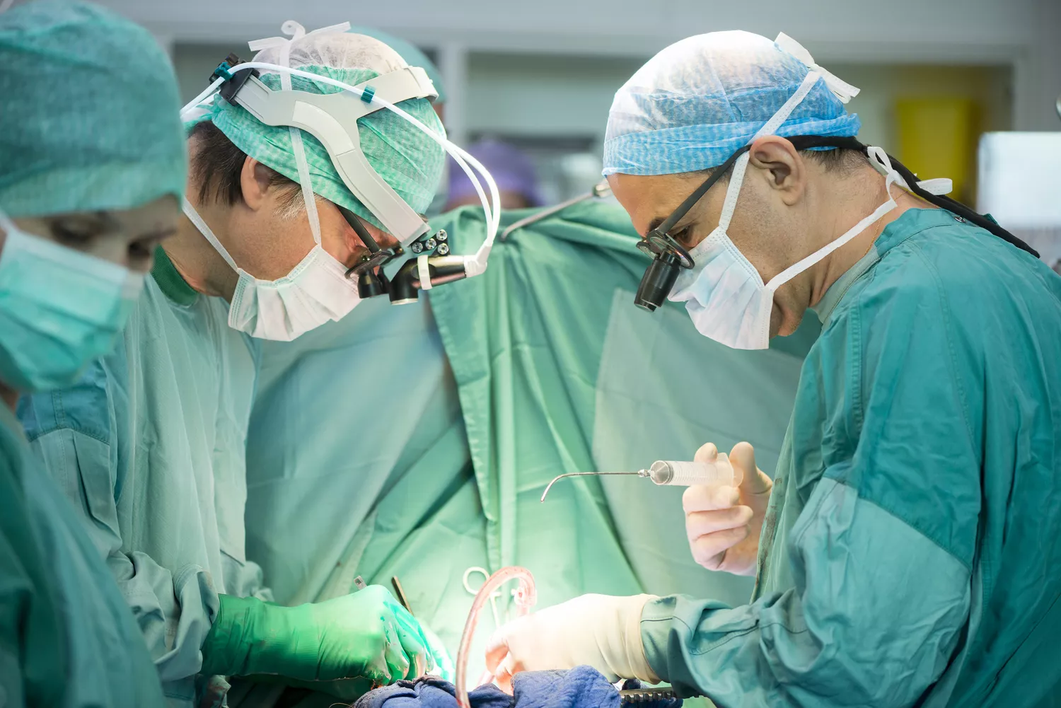
JazzIRT / Getty Images
What Is Angiography Used For?
Angiography may be used to:1
Locate blockages in the lung (pulmonary), heart (coronary), brain (cerebral), and other smaller blood vessels
Find sites of internal bleeding, or hemorrhage
Find aneurysms, or abnormal dilation of blood vessels, which may cause major health problems
Observe abnormal blood flow caused by stenosis, or narrowing of the blood vessels
Identify problems with the structure of the heart
Locate internal bleeding or other obstructions that should be removed
Abnormal blood flow affects the organs supplied by the vessels, and may increase the risk for chest pain (angina), heart attack, stroke, and other disorders.1
Angiography may also be used to deliver treatment. For example, angioplasty may be done to remove blockages and open up narrowed arteries. It is also possible to deploy fixed dilators called stents to widen arteries and coil or seal off aneurysms as part of an angiogram procedure.2
Before an Angiogram
Prior to angiography, the healthcare provider who is conducting the procedure will likely take a thorough history and do a physical examination. They will also go over the purpose, risks, and benefits of the angiogram.
This is an excellent opportunity to ask any questions that might come up.
Your healthcare provider may want to use a more advanced imaging technique like angiography for diagnosis if initial non-invasive testing has been inconclusive.
Timing
It will be important to arrive prior to the testing for the intake process. This may involve completing paperwork, changing into a hospital gown, and having intravenous access placed.
Prior to the procedure, you will be transferred into the suite where the angiogram is performed. Depending on the intervention, the angiogram procedure may last more than an hour. Recovery after may add several additional hours.
Location
Angiography takes place in the catheterization lab or “cath lab” of a hospital or medical center. This sterile room has X-ray equipment, viewing monitors, and an examination table where you will lie still during the procedure.
What to Wear
Individuals undergoing an angiogram will disrobe and change into a hospital gown.
Food and Drink
To prepare for angiography, it is important to avoid eating in the eight hours leading up to the procedure. Drinking clear liquids until two hours before the procedure will help keep blood vessels open, flexible, and more easily accessible.
Cost and Health Insurance
The procedure may require a prior authorization process to ensure insurance coverage. Deductibles and co-payments may add to the out-of-pocket expense. Without insurance, the procedure could easily cost thousands of dollars.
What to Bring
It will be important to bring identification and health insurance information to the procedure. Comfortable, loose-fitting clothing is recommended to wear home. You will also need someone to drive you home after the angiogram is completed.
During Angiography
The healthcare provider, often a specialist in either cardiology or neurology, will lead a team that may include nursing staff as well as other providers, potentially including an anesthesiologist.
Pre-Test
Local or general anesthetics are given to sedate you and numb the access point.
In most cases, you will be awake during an angiogram, however, medications will be given to help you feel relaxed.1 In some cases, like if a child is undergoing angiography, sedation may be heavier so they sleep during the procedure.
Throughout the Test
The angiography procedure involves several steps:
After creating a small incision, a sheath is inserted into the blood vessel which allows for the insertion of the guidewire and catheter, as well as the injection of contrast medications.
The guidewire is visible with X-ray and can be tracked as it progresses through the circulatory system.
Once the guidewire is in place, a catheter is inserted over the guidewire and threaded to the target blood vessel where it feeds the contrast agent into the bloodstream.
Is an Angiogram Painful?
Throughout this process there may be mild stinging, pressure, or discomfort at the insertion site.
Coronary angiography, which is done to see the heart's blood vessels, is slightly different. It involves the following:
A local anesthetic is used to numb the area where the catheter is inserted—typically the brachial artery in the forearm or the femoral artery at the groin. A general anesthetic may be used if high levels of anxiety or discomfort would disrupt the procedure or emotional well-being.
A guidewire and catheter are inserted and guided through the arterial system until they reach the major coronary arteries.
Throughout the procedure, the catheter may be relocated to make images of other parts of the arterial system or to directly image the interior of the heart.
If conscious, you may be asked to take a breath in and hold it at certain points during the procedure. There may be sensations of heat or discomfort as the contrast agent enters the heart directly.
Angiography may take an hour or more, depending on what is required.
Post-Test
As the procedure ends, the catheter will be removed and a healthcare provider will apply pressure to the access site and monitor to ensure bleeding is not present. Often you will remain lying flat for a specified period of time.
Higher-risk angiograms, such as when the femoral artery is accessed, may require you to stay at the hospital for a few hours of bed rest and observation. You should not drive home.
After Angiography
Some people feel back to normal within a day or two. For the day after the procedure, it can be helpful to have someone around to monitor for any issues that may develop. They may need to help prepare food or administer medications. If there is a serious problem, it may be necessary to contact the healthcare provider and get emergency medical assistance.
For 24 hours following angiography, you should not:
Drink alcohol
Smoke
Perform tasks that require coordination (such as operating vehicles or heavy machinery)
For three days following angiography, it is important to avoid:
Exercise
Sexual intercourse
Submersion in water (such as in a bath or swimming pool)
Engaging in certain activities within days following the procedure can reopen the access wound and increases your risk for side effects.
Managing Side Effects
If bleeding continues at the entry site, stay relaxed, apply direct pressure, and contact your healthcare provider as soon as possible.
If your chest hurts after an angiogram, call your healthcare provider. While you may notice some bruising, let your healthcare provider know if you have any pain, swelling, numbness, or fever.1
What Are the Risks of Angiography?
As with any medical procedure, there is the possibility of side effects or complications. These may be more likely if there are procedural mistakes, allergies, or coexisting medical conditions. Major complications are exceedingly rare and almost never fatal, so there is no specific set of risk factors to prevent someone from having an angiogram.3
General risks of angiograms include:
Bruising
Bleeding
Pain
Infection
Injury to blood vessels
However, informing a healthcare provider about certain factors, such as allergies, may lead to changes in the preparation and execution of the procedure, which may help to decrease risk. Technological advances have also decreased the likelihood of mechanical damage caused by the equipment and poor physiological responses to the substances used for pain relief and imaging.
Allergic responses can occur due to a number of substances used in the procedure, and asthma or the use of beta-adrenergic blockers increase the likelihood of a serious allergic response. The mechanical movements of the instruments during the procedure can also cause problems like bleeding and clotting, which may, in turn, induce more serious complications like:
Bleeding
Aneurysm
Stroke
Heart attack
Death
The risk of the procedure is always weighed against the potential benefit, which is often very high.
Local Anesthetic Reactions
The most likely reactions caused by local anesthetics, or the preservatives they may contain, are skin irritation at the injection site or fainting. Restriction of breathing (anaphylaxis) can occur, but this is rare. A history of allergic responses to local anesthetics or preservatives could warrant skin testing before an angiography is performed or substituting the use of preservative-free anesthetics.
Allergies or Toxicity
Some people may be at risk of allergies or complications from general anesthetics, contrast media, or heparin use during the procedure.
General Anesthetics
Though general anesthetics are rarely needed to perform angiography, some risks are involved if they are utilized. While an anaphylactic allergic response is unlikely with conscious sedation, general anesthetics can affect the function of the heart if given at an improper dose.
The goal of general anesthesia in angiography is to limit sensation, rather than to make a person unconscious. However, loss of sensation could mask the recognition of some kinds of complications.
As with any surgical procedure, vital signs such as heart rate, heart rhythm, blood pressure, and blood oxygen level are monitored to identify any abnormal changes in heart or lung function.
In situations of an anesthetic overdose, reversal agents may be provided to restore normal organ function. A severe anaphylactic response may be treated with epinephrine, corticosteroids, high-flow oxygen delivered via a mask, and even intubation and ventilation until the reaction subsides.
Contrast Media
Though many types of contrast media have become available which drastically decrease sensitivity reactions, it may cause anaphylaxis and chemotoxicity. Contrast media can cause constriction of the throat through the release of histamines or induce allergic responses to iodine.
Chemotoxicity can result due to the interaction of the contrast media and blood. Minor side effects include:
Warmth
Pain
Tightness
Nausea
Vomiting
Major side effects include:
Low blood pressure (hypotension)
Slowed heart rate (bradycardia)
Fluid in the lungs (pulmonary congestion)
In addition, contrast-induced nephropathy (CIN) is another danger that can affect people with vulnerable kidneys. Newer contrast media may reduce the risk. Decreasing the volume of contrast media administered and promoting hydration with intravenous fluids before, during, and after the procedure may also help.
Heparin-Induced Thrombocytopenia (HIT)
Heparin is a blood thinner used during angiography. In susceptible individuals, exposure to heparin may cause an amplified immune system response that activates platelets and leads to clotting and inflammation in blood vessels.4
This may cause blood clots to form via thrombosis. As the supply of platelets in the blood is consumed, there may be an increased risk of bleeding (and possible hemorrhage). Treatment is possible and the platelet counts may be monitored to ensure normalization.
Physical Disruptions
A physical disruption could be an injury, irritation, or infection that happens during the procedure. These include:
Local Vascular Injury
One risk is bleeding caused by damage to the blood vessels as the catheter is inserted and moved internally. Reduction of catheter size and increased use of fluoroscopy (real-time X-ray visualization to guide the wire) have been useful in finding and inserting into target blood vessels correctly without causing damage. Nevertheless, the risk still exists and may be made worse by factors that decrease clotting ability.
Hematoma
When the catheter sheath is removed at the end of surgery, blood can pool outside of the peripheral artery at the point of insertion, forming a mass called a hematoma. Hematomas most notably occur near the femoral artery. They are not generally harmful, but larger ones can block blood vessels (potentially leading to thrombosis) or compress nearby nerves.
The angiography procedure includes steps taken to reduce hematoma risks. Post-surgery, your healthcare provider applies pressure to the sheath site to keep large hematomas from forming. Likewise, resting after surgery limits the risk of forming hematomas.
If a hematoma causes dangerous blood loss, a second angiogram may be performed to identify and repair the damaged blood vessel.
False Aneurysm
A false aneurysm (called a pseudoaneurysm) can occur when a smaller artery is accidentally catheterized. The mismatch of size can damage the wall of the blood vessel and cause the subsequent formation of an aneurysm, an extension outside of the normal blood vessel. Most pseudoaneurysms can be spotted with an ultrasound and then treated with an injection of thrombin which stops blood from flowing into the aneurysm.
Arteriovenous Fistula (AVF)
An arteriovenous fistula (AVF) may form when an artery and vein are penetrated near each other and form a connection, allowing the higher arterial pressure to enter into the vein. Most fistulas should be monitored but will close off with time.
Dissection
Dissection, or cutting, of the femoral or iliac artery during sheath placement is very rare but risks limb loss or even death if untreated. A stent may need to be placed to restore normal blood flow to the limb and allow the damaged artery to heal.
Thrombosis and Embolism
While the sheath and catheter are in place, they can disrupt flow through the blood vessel. Blockage can occur, particularly in people with smaller blood vessel size, arterial disease, or diabetes. A clot, or thrombus, may form. The risk may be reduced by regularly flushing the sheath and using anticoagulants during longer procedures.
A blood clot that travels along the bloodstream to cause damage at another site, called an embolism, may result in a stroke as well as numbness or pain affecting the limbs, hands, or feet. This may need to be treated with surgery to remove the clot (called thrombectomy).
Cholesterol Emboli
Physical disruption of cholesterol deposited along the lining of blood vessels can lead to an embolism. These cholesterol plaques commonly narrow blood vessels in atherosclerosis. Symptomatic occurrences of cholesterol emboli associated with an angiogram are rare.
Findings may include discoloration of an extremity or splotchy, purple patterns in the skin (known as livedo reticularis). Risk factors include age, repeated vascular procedures, and elevated amounts of inflammation-driven C-reactive protein.
Bradycardia
Bradycardia, or low heart rate, can be caused by irritation or blockage as the catheter nears the heart. When this occurs, an affected individual may begin to feel nauseous, sweat, or yawn. The healthcare provider will adjust the catheter position and monitor vital signs. If the catheter caused a blockage affecting heart function, a forceful cough or the intravenous administration of atropine may help to recover the normal heart rate.
Tachycardia
The opposite problem, tachycardia (a high heart rate), can also be caused by irritation from the catheter. It is usually immediately reversible by pulling back the catheter. If it persists and leads to an unstable blood pressure, this may require defibrillation.
Infection
The risk of infection in the setting of an angiogram is very low, but people who have a fever or other symptoms may require medical treatment.
Signs of Infection After Surgery
Significant Morbidity and Mortality
In rare cases, complications from angiography can lead to stroke or heart attack, which can be fatal.
Stroke
Hypertension, diabetes, prior strokes, abnormal kidney function, and emergency angiography can increase the risk of a stroke occurring during the procedure. An embolus that travels to the brain may occur when thrombosis occurs near the catheter or when plaque is dislodged. Stroke occurs in less than 1% of people with risk factors.3
Heart Attack (Myocardial Infarction)
Heart attack can occur during angiography, but this happens in under 0.2% of angiograms. It is more likely to occur in longer, more complicated procedures.3
Death
Unfortunately, death may also occur due to angiography in rare circumstances. Recent heart attacks, left main coronary artery disease, aortic stenosis, increased age, and poor kidney function are the main risk factors that increase the chance of death. Mortality occurs in less than 0.1% of angiograms, affecting 1 in 1000 people undergoing the procedure, but this outcome is more likely in those with known risk factors.3
Interpreting Results
Often an angiogram is performed with both a diagnostic portion, to better visualize the nature of the problem, and a treatment portion, in which an intervention immediately corrects the underlying problem. Unlike other tests, it is often unnecessary to gather information to review and be used at a later date.
Due to the nature of the procedure, it is best to intervene promptly during the time that the individual is both medicated and the arterial access exists. Prior to the angiogram, the healthcare provider will outline the likely findings as well as how any abnormalities that are identified may be corrected before the conclusion of the procedure.
Follow-Up
You'll need a follow-up appointment with your healthcare provider in the weeks after the angiogram. This allows you to discuss the results of the intervention and whether your symptoms have improved. In rare cases, an angiogram may be repeated for further evaluation or intervention.
Types of Angiography
A healthcare provider may want to perform different types of angiography to identify the source of the problem, make a diagnosis, and plan the next steps in the treatment, like surgery, medication, or behavioral changes.
Coronary Angiography
The coronary arteries supply blood flow to the heart and are vital to its function. Coronary angiography may be used if you are experiencing:
Chest pain
Change in heart rate
Change in blood pressure
Unexplained pain affecting the jaw, neck, or arm
Serious concerns, such as abnormal heart rhythms, heart attack, or congestive heart failure, which is when the heart doesn't pump enough blood for the body's needs
Congenital heart defect, a malformation present since birth
Aortic stenosis, or a type of heart valve condition
Heart valve disease, which describes one or more valves not functioning properly
Chest injury
Cerebral Angiography
It is also possible to image the blood vessels to the brain with cerebral angiography. Additional imaging techniques may be used with the procedure to enhance the visualization.
Cerebral angiography may be used to:
Treat the narrowing that contributes to transient ischemic attacks, also called mini strokes, or those at risk for a stroke
Treat a stroke-related clot
Seal off cerebral aneurysms, abnormal dilation or bulging of blood vessels, which are prone to rupture and secondary hemorrhage
Microangiography
Microangiography may be used to image the smaller blood vessels supplying other organs, particularly to address localized bleeding. It may also be useful in detecting and treating cancerous tumors since fast growing tumors contain a lot of blood vessels.
Summary
An angiogram is an effective procedure to diagnose and treat disorders that commonly affect the blood supply of the heart and brain. The procedure involves injecting contrast substances into the blood to better show how it flows. Risks of injury from angiography are generally slight, but complications, such as an allergic response or kidney issues, are possible.


Post comments