
by Elana Gotkine
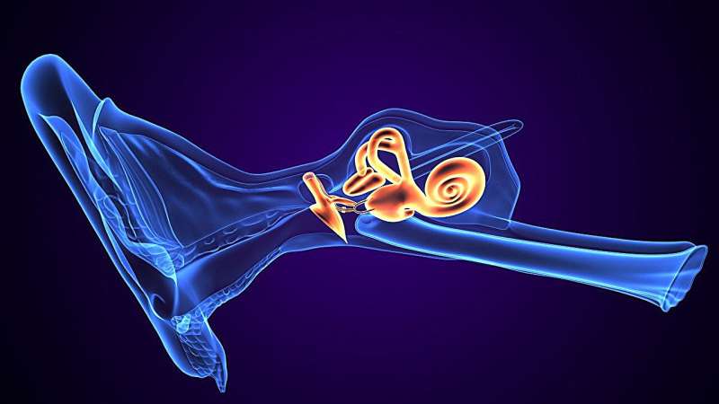
Distension of the membranous labyrinth is seen in histologic specimens from deceased patients with symptoms consistent with Meniere disease and in magnetic resonance imaging (MRI) from patients exhibiting classic Meniere disease symptoms, according to a study published online June 13 in the Ear, Nose & Throat Journal.
Eugene D. Ark, from the College of Medicine at the University of Illinois-Chicago, and colleagues explored the distended membranous labyrinth through a comparison of early temporal bone studies with contemporary magnetic resonance imaging techniques.
Histopathological photomicrographs from deceased patients with symptoms consistent with Meniere disease were reviewed, and MRI sequences were captured from patients exhibiting classic Meniere disease symptoms four hours after gadolinium administration.
The researchers found that in patients with Meniere disease, both histopathologic examination and MRI imaging revealed consistent distention of the saccule, utricle, and scala media. The correlation between postmortem histological findings and MRI evidence of distension in living patients was successfully demonstrated.
"This study's overall aim is to raise public awareness that these neglected collections are valuable and should be curated," the authors write. "This article demonstrates the correlation of traditional histopathologic findings with the MRI-demonstrated distention of the endolymphatic spaces."
More information: Eugene D. Ark et al, Four-Hour-Delayed Gadolinium 3D REAL IR and SPACE FLAIR MRI Correlated to Meniere Disease Histology, Ear, Nose & Throat Journal (2024). DOI: 10.1177/01455613241261461
© 2024 HealthDay. All rights reserved.


Post comments