Driving and dementia—assessing safe driving in high-risk older adults
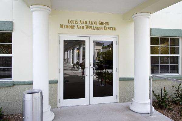
by Florida Atlantic UniversityFAU's Louis and Anne Green Memory and Wellness Center operated by the Christine E. Lynn College of Nursing provides a comprehensive driving evaluation that inclu
Updated on: June 04,2024
Driving and dementia—assessing safe driving in high-risk older adults
by Florida Atlantic UniversityFAU's Louis and Anne Green Memory and Wellness Center operated by the Christine E. Lynn College of Nursing provides a comprehensive driving evaluation that inclu
Updated on:June 04,2024