
byTechnical University Munich
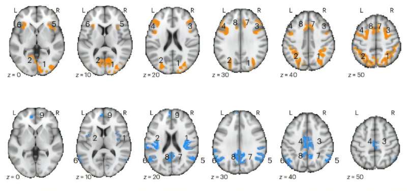
Regional clusters (i; designated by numbers) of positive (top) and negative (bottom) ∆BOLD (PLS group results, thresholded at BSR > ±3). Credit:Nature Neuroscience(2025). DOI: 10.1038/s41593-025-02132-9
For almost three decades, functional magnetic resonance imaging (fMRI) has been one of the main tools in brain research. Yet a new studypublishedinNature Neurosciencefundamentally challenges the way fMRI data have so far been interpreted with regard to neuronal activity.
According to the findings, there is no generally valid coupling between the oxygen content measured by MRI and neuronal activity.
Researchers at the Technical University of Munich (TUM) and the Friedrich-Alexander-University Erlangen-Nuremberg (FAU) have found that an increased fMRI signal is associated with reduced brain activity in around 40% of cases. At the same time, they observed decreased fMRI signals in regions with elevated activity.
First author Dr. Samira Epp emphasizes, "This contradicts the long-standing assumption that increased brain activity is always accompanied by an increased blood flow to meet higher oxygen demand. Since tens of thousands of fMRI studies worldwide are based on this assumption, our results could lead to opposite interpretations in many of them."
PD Dr. Valentin Riedl, now Professor at FAU, and his colleague Epp examined more than 40 healthy participants during their time at TUM. Each was given several experimental tasks—such as mental arithmetic or autobiographical memory recall—which are known to produce predictable fMRI signal changes in distributed brain regions.
During these experiments, the researchers simultaneously measured theactual oxygen consumptionusing a novel quantitative MRI technique.
Depending on the task and the brain region, the physiological results varied. Increased oxygen consumption—for instance in areas involved in calculation—did not coincide with the expected rise in blood flow.
Instead, thequantitative analysesshowed that these regions met their additional energy demand by extracting more oxygen from the unchanged blood supply. Thus, they used the oxygen available in the blood more efficiently without requiring greater perfusion.
According to Riedl, these insights also affect the interpretation of research findings in brain disorders. "Many fMRI studies on psychiatric or neurological diseases—from depression to Alzheimer's—interpret changes in blood flow as a reliable signal of neuronal under- or over-activation.
"Given the limited validity of such measurements, this must now be reassessed. Especially in patient groups withvascular changes—for instance, due to aging or vascular disease—the measured values may primarily reflect vascular differences rather than neuronal deficits."
Previous animal studies already point in this direction.
The researchers therefore propose complementing the conventional MRI approach with quantitative measurements. In the long term, this combination could form the basis forenergy-based brain models: rather than showing activation maps that depend on assumptions about blood flow, future analyses could display values indicating how much oxygen—and therefore energy—is actually consumed for information processing.
This opens new perspectives for examining aging, psychiatric, or neurodegenerative diseases in terms of absolute changes in energy metabolism—and for understanding them more accurately.
More information Samira M. Epp et al, BOLD signal changes can oppose oxygen metabolism across the human cortex, Nature Neuroscience (2025). DOI: 10.1038/s41593-025-02132-9 Journal information: Nature Neuroscience


Post comments