
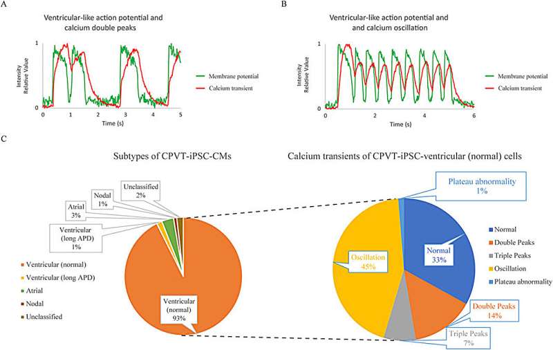
Analysis of cardiomyocytes differentiated from type 1 catecholaminergic polymorphic ventricular tachycardia (CPVT) patient-derived human induced pluripotent stem cells (CPVT1 hiPSC-CMs) using the system to record action potentials (APs) and Ca2+ transients simultaneously. Credit: Frontiers in Physiology (2025). DOI: 10.3389/fphys.2025.1579815
Researchers have developed a novel method for simultaneously recording action potentials (APs)—temporary changes in electrical potential when cells become excited—and calcium transients—calcium fluctuations that drive muscle contraction—in single cardiomyocytes (heart muscle cells) derived from iPS cells.
The study, led by Associate Professor Yoshinori Yoshida, Department of Cell Growth and Differentiation at Kyoto University in collaboration with Takeda Pharmaceutical Company, was published in Frontiers in Physiology.
Catecholaminergic polymorphic ventricular tachycardia (CPVT) is a rare but life-threatening inherited arrhythmia characterized by abnormal calcium handling in cardiomyocytes. While human iPS cell-derived cardiomyocytes have been widely used to model CPVT and test drug responses, most studies have focused independently on either action potentials or calcium transients.
This separation limits the ability to fully understand the interplay between electrical and calcium signaling abnormalities that drive arrhythmias. As such, a method enabling simultaneous, high-resolution recording of both APs and calcium transients in single cells is essential for more accurate disease modeling and drug evaluation.
To this end, the team used a combination of the membrane potential dye FluoVolt and the newly developed calcium indicator Calbryte 590 AM. This dual-dye approach achieved a higher signal-to-noise ratio than conventional methods, allowing for long-term, simultaneous imaging of AP and calcium waveforms in single cells. Furthermore, the researchers optimized light intensity and filter settings to minimize phototoxicity and maximize detection accuracy.
Using this system, the team analyzed cardiomyocytes derived from a CPVT type 1 (RyR2-I4587V) patient's iPS cells. They found two-thirds of cardiomyocytes resembling ventricular cells to exhibit abnormal calcium transients such as calcium double peaks, triple peaks, and oscillations significantly more than healthy control cells, consistent with known CPVT pathophysiology.
The researchers then tested the effects of several drugs on these abnormal cells. Carvedilol, a nonselective β-blocker, improved calcium abnormalities at 1-3 µM, though higher concentrations induced atrial-like APs or asystole. Flecainide, a class 1C antiarrhythmic, showed moderate efficacy at 1-10 µM, aligning with clinical observations.
JTV519 (K201), a RyR2 modulator, significantly improved calcium abnormalities at 3 µM but also altered AP morphology. Notably, the CaMKII inhibitor KN-93 demonstrated the highest efficacy, normalizing calcium transients in 93% of cells at 1 µM without affecting APs.
By enabling simultaneous monitoring of electrical and calcium dynamics in patient-derived cardiomyocytes, the method provides a more comprehensive and physiologically relevant platform for evaluating drug responses. It supports the development of safer, more effective therapies tailored to individual patients and may help identify new treatment strategies for arrhythmias that are difficult to manage with current options.
Importantly, this is one of the first methods to achieve stable, long-term dual imaging in single human iPS cell-derived cardiomyocytes. Beyond CPVT, it could be adapted to study other cardiac disorders and drug-induced arrhythmias and may serve as a foundation for future precision cardiology tools.
More information: Tadashi Takaki et al, Simultaneous optical recording of action potentials and calcium transients in cardiac single cells differentiated from type 1 CPVT-iPS cells, Frontiers in Physiology (2025). DOI: 10.3389/fphys.2025.1579815 Journal information: Frontiers in Physiology


Post comments