
Clinical Pearl Series Edited from Yale-G First Aid: Crush USMLE Step 2CK & Step 3 by Yale Gong, MD, Sr. Medical Advisor at www.medicine.net (Copyrighted)
Cardiovascular disease has been the leading cause of death worldwide for decades, with its high incidence clearly linked to modern lifestyles—excessive nutrition, insufficient exercise, excessive stress, and exposure to tobacco, alcohol, and chemical toxins. Therefore, routine health checkups, health counseling, and lifestyle adjustments can significantly reduce its incidence and mortality rates.
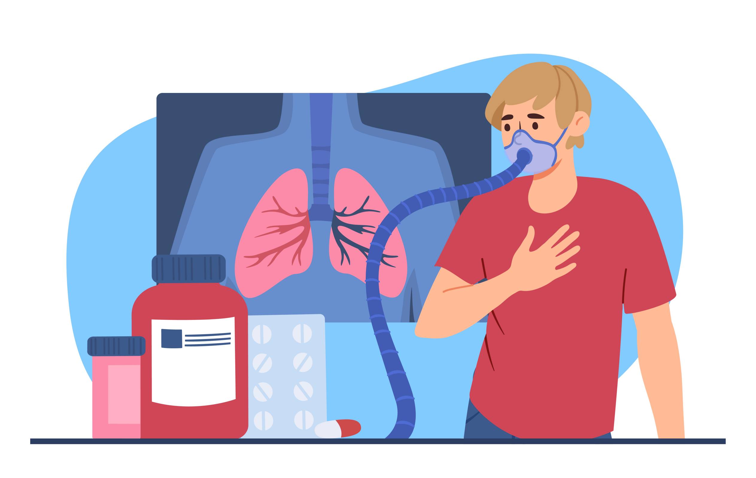
Chest pain or upper abdominal pain is one of the most common symptoms in the majority of cardiovascular diseases, respiratory diseases, and certain upper abdominal conditions. Therefore, mastering the key points of differential diagnosis is crucial for early, timely, and accurate diagnosis and treatment, ultimately saving lives!
I. Cardiac Conditions
Angina
It mostly refers to “stable angina”, a paroxysmal chest pain resulting from cardiac ischemia mostly caused by atherosclerotic coronary arteries. Stable angina is the type when the chest pain is precipitated by predictable factors (exercise, exertion, etc.). Unstable angina is angina that occurs at any time and carries more risk for myocardial infarction (considering as a pre-MI condition).
Major risk factors: Age (male > 45, female > 55 y/a), male gender, smoking, hypertension, diabetes, heredity (including race, family history < 55 y/a), atherosclerosis, dyslipidemia (LDL > 200 mg/dL, HDL < 40 mg/dL), physical inactivity, obesity, stress, excess alcohol use, and postmenopausal women.
Clinical features:
(1) Pressing or squeezing chest pain radiating to the left jaw or arms, lasting for 15 sec—15 min. (2) Precipitating factors: exertion, anxiety, meals, and coldness. 3Pain relief: Nitroglycerin, resting. (3) ECG: ST-T depression. Treatment: (1) Nitrates; (2) Beta1 receptor blockers (Atenolol, metoprolol). (3) Ca-channel blockers (verapamil, etc.). (4) Unstable angina: Hospitalize and treat the patient with aggressive aspirin, nitrates, beta-R blockers, and heparin, and be ready for revascularization.
Myocardial infarction (MI)
MI is ischemic myocardial necrosis as a result of an abrupt reduction in the coronary blood flow to a segment of myocardium, usually due to a thrombotic occlusion of a coronary artery previously narrowed by atherosclerosis. MI is associated with a 30% mortality rate and 50% pre-hospital deaths. Emergency management at initial symptoms is life-saving! Risk factors: Same as angina.
Clinical features:
(1) Characteristic symptoms: severe, crushing, prolonged chest pain more severe than angina; associated with dyspnea, anxiety, diaphoresis, nausea, vomiting, weakness, low fever, sense of impending doom, and syncope (in elderly). Painless and atypical MI can be up to 1/3 cases and more likely in postoperative or diabetic patients and the elderly. Sudden cardiac death can occur due to ventricular fibrillation (V-fib).
(2) Cardiogenic shock signs: seen with > 40% cases of MI—BP decrease, S3 gallop, and rales.
(3) ECG: peaked T waves (early), ST-segment elevation (transmural infarct) or depression (subendocardial lesion), or Q waves (necrosis, late).
Treatment:
(1) “ABC” first—airway, breathing, and circulation. Supplemental oxygen. (2) Treat sustained ventricular arrhythmia or heart failure rapidly. (3) Beta1-R blockers (metoprolol): shown reduced post-MI mortality rate. (4) Nitrates. (5) Antiplatelet therapy: Aspirin (PO).
(6) Thrombolytic therapy. (7) Analgesics: IV opiates (morphine) are important to relieve pain, to supply relaxation and sedation, and to alleviate CVS and respiratory stress effectively. (8) ACE inhibitors (Angiotensin-converting enzyme inhibitors, ACE-I): It has shown reduced post-MI mortality. (9) Hypolipidemic therapy: Atorvastatin should also be started early and before percutaneous coronary intervention (PCI). (10) Coronary angiography and angioplasty.
Myocarditis
It’s acute or chronic infection or inflammation of the myocardial cells, leading to reduced cardiac contractility and output, and possibly heart failure. It’s usually preceded by an upper respiratory viral infection (fever, sore throat) and associated with a vague chest pain and increased creatine kinase (CK). ECG shows abnormal conduction or Q waves. Be cautious that a severe type of viral myocarditis may progress rapidly to myocardial infarction with a senior age or direct heart failure with a young age.
Treatment: Supportive and symptomatic therapy is the mainstay of therapy. anti-inflammatory drugs and alcohol should be avoided.
Pericarditis
It’s the inflammation of the pericardial lining around the heart. It may be preceded by a viral illness. Chest pain is sharp, pleuritic, and positional—worse with lying down and relieved by sitting up. Pericardial rub often exists. ECG usually shows diffuse ST elevation without Q waves. CK is mostly normal. It responds well to anti-inflammatory drugs.
Mitral valve prolapse
A common congenital valvular abnormality characterized by transient non-angina chest pain with a typical midsystolic click murmur. Echocardiography is the best method for diagnosis. Most cases are asymptomatic and not in need of treatment.
II. Pulmonary Conditions
Pneumonia
Typically caused by bacterial infection in the lungs, presenting with moderate chest pain accompanied by high fever, chills, cough, fatigue, and sputum production. May or may not be accompanied by hemoptysis or cyanosis of the lips. Initial evaluation involves a chest X-ray; if complications or tumors are suspected, a CT scan is performed. Treatment involves targeted antibiotics and supportive care.
Pneumothorax
It is the accumulation of air in the pleural cavity between the lungs and the chest wall. Typical manifestations include sudden, sharp, pleuritic chest pain and dyspnea; absent breath sounds; mediastinum shifted to the opposite site—suspect of tension pneumothorax—requiring urgent intercostal needle puncture. Non-tension pneumothorax can wait for CXR confirmation and natural relief.
III. Vascular Conditions
Aortic dissection
It involves tearing of the inner layer of the aorta, allowing blood to enter the layers of the vessel wall and form a “false lumen.” It is classified into types A and B. Typical symptoms include severe, sharp, tearing chest pain radiating to the back; absent pulse, unequal blood pressure in both arms, or signs of aortic valve insufficiency; neurological signs; and a widened mediastinum on chest X-ray. If the dissection extends to the coronary arteries, myocardial infarction may occur. Diagnosis is confirmed via transesophageal echocardiography (TEE), CT scan, or aortic angiography. An aortic aneurysm is a localized dilation and bulging of the aorta, exceeding 1.5 times the normal vessel diameter. Treatment: Type A dissection and ruptured or impending aortic aneurysms require emergency fluid and blood transfusion followed by life-saving surgery.
Pulmonary (artery) embolism (PE)
Typically occurring after prolonged sitting during flying or inactivity within 3-5 days post-surgery, deep vein thrombosis in the lower extremities forms clots that travel back to the right atrium and ventricle, obstructing the pulmonary artery. This causes sudden onset of chest pain, shortness of breath, tachycardia, coughing, and hypoxemia (such as cyanosis). Chest pain is typically pleuritic but may mimic angina. CT pulmonary angiography has replaced V/Q scans as the preferred diagnostic method.
Pulmonary hypertension
This is a pathological state characterized by elevated pulmonary arterial pressure due to various etiologies. It is associated with a history of chronic cardiovascular or pulmonary disease; dull chest pain accompanied by symptoms and signs of chronic right ventricular failure, primarily manifested as persistent lower limb edema, hepatomegaly, and jugular venous distension, with symptoms progressively worsening. Treat primary disease.
IV. Musculoskeletal Conditions
Costochondritis
It is classified into nonspecific costochondritis and infectious costochondritis, representing the most common cause of chest pain, particularly in younger individuals. Symptoms typically manifest as localized, spontaneous pain and tenderness that worsens during inhalation, coughing, sneezing, or chest palpation, with symptoms often recurring. Electrocardiograms (ECGs) are normal. Treatment involving rest combined with common anti-inflammatory pain relievers (such as ibuprofen) proves effective. ECG abnormalities typically indicate cardiac disease, making ECG a necessary diagnostic tool for individuals with cardiovascular risk factors.
V. Gastrointestinal Conditions
Gatric diseases
Gastroesophageal reflux disease:Gastroesophageal Reflux Disease: A condition caused by multiple pathological mechanisms leading to the reflux of stomach contents into the esophagus. Typical symptoms include burning chest pain, acid regurgitation, a sour taste, and difficulty swallowing, which may be relieved by antacids. Persistent reflux may cause esophageal mucosal damage and chronic inflammation, potentially leading to esophageal stricture and upper gastrointestinal bleeding. Treatment involves avoiding triggers (such as alcohol and high-fat foods) and using medications that reduce stomach acid.
Gastric Spasm: Refers to excessive contraction of the smooth muscles in the stomach wall, presenting as sudden, severe upper abdominal pain accompanied by nausea, vomiting, and other symptoms. Primary causes include improper diet (such as raw, cold, or spicy foods), emotional factors, and abdominal exposure to cold. Commonly seen in conditions like acute/chronic gastritis and gastric or duodenal ulcers. Relief: Apply heat and massage, take pain medication, and adjust diet. Treat the underlying condition.
Peptic Ulcer Disease: Primarily refers to chronic gastric and duodenal ulcers, with common symptoms including upper abdominal pain before or after meals. Causes include Helicobacter pylori infection and prolonged use of anti-inflammatory pain medications. Diagnosis relies on endoscopy and H. pylori testing. Treatment involves antacids, antibiotic therapy to eradicate H. pylori, and avoiding anti-inflammatory drugs.
Acute pancreatitis
It’s an inflammation of the pancreas caused by multiple factors, with common triggers including gallstones, chronic alcohol abuse, and hyperlipidemia. It typically presents as persistent, severe upper abdominal pain radiating to the back following a heavy meal, accompanied by nausea, vomiting, fever, and elevated serum amylase and lipase levels. Hospitalization is usually required, involving fasting and gastrointestinal decompression.
Cholangitis/Gallstones
Right upper quadrant (RUQ) pain typically occurs after greasy meals, accompanied by tenderness, nausea, vomiting, and/or jaundice. This condition predominantly affects middle-aged obese women, and B-mode ultrasound aids in diagnosis. Delayed treatment may lead to serious complications such as biliary obstruction or gallbladder perforation. Comprehensive management should be tailored to the specific circumstances.
Hiatal hernia
This refers to the stomach or intestines protruding into the chest cavity through the esophageal hiatus in the diaphragm. This condition may lead to complications such as reflux esophagitis, and long-term untreated cases may increase the risk of esophageal cancer. Common symptoms include burning pain in the chest and upper abdomen, regurgitation of food, acid reflux, nausea, and vomiting. Severe cases may present with difficulty swallowing and breathing. Treatment options include medication and surgery; antacids may alleviate symptoms.


Post comments