
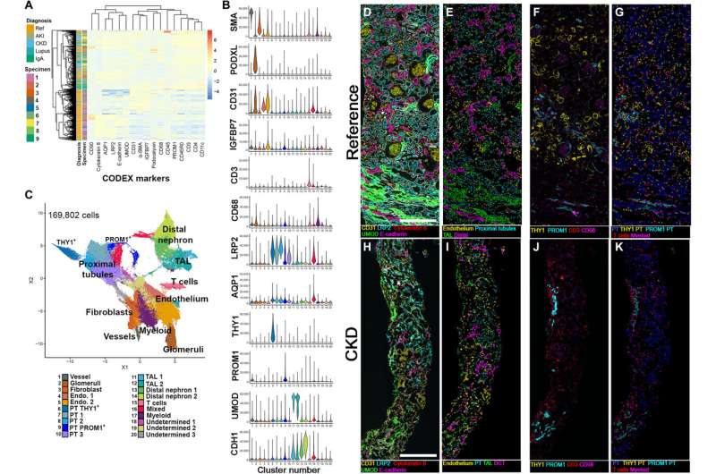
Cell classification based on CODEX imaging in reference and disease specimens. Reference and disease specimens were stained and imaged using CODEX. Credit: Science Advances (2025). DOI: 10.1126/sciadv.adv8918
A research team led by Indiana University School of Medicine physician scientists has made significant progress in mapping kidney cells that may one day allow for more accurate disease diagnosis.
This work, published in Science Advances, is part of a greater IU School of Medicine-led collaborative effort to identify and characterize the many diverse cell types in the human kidney.
"Understanding of the spatial organization of the kidney cell populations and their interaction within molecular neighborhoods will allow us to define a better timeline of kidney disease based on molecular staging, " said Tarek M. Ashkar (El-Achkar), MD, a corresponding author and the Terence P. Kahn Professor of Nephrology at IU School of Medicine.
"This, in turn, will uncover better biomarkers and targets for therapy that reflect the correct pathology at the right stage of the disease, " Ashkar added. "This will lead to treating the right patient at the right time with the right medicine."
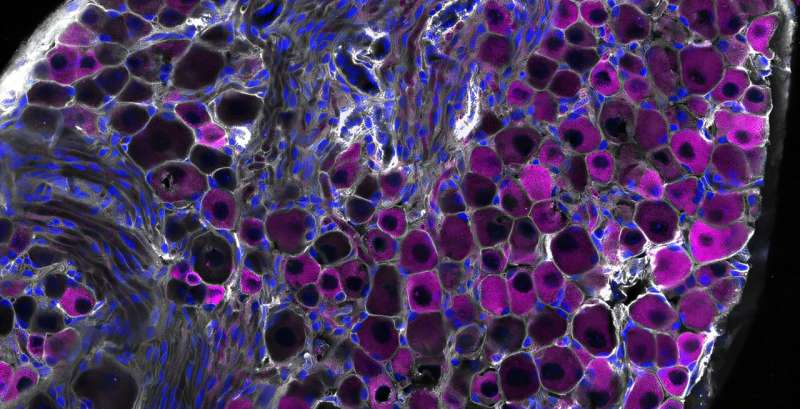
The team identified two subpopulations of proximal tubule cells: one regenerative type plentiful in healthy kidneys, and another featuring a genetic marker that signals disease. By measuring the number of each type of cell in kidney tissue, researchers can more accurately map the level of disease present.
Kidney diseases are a leading cause of death in the United States, according to the U.S. Centers for Disease Control and Prevention. About 14% of adults suffer from chronic kidney disease, and many do not know they have it. Treatment for kidney failure is either dialysis or transplant, both of which have a major impact on the patient's quality of life.
Kidney cells are much more complex than previously believed, Ashkar said. One larger goal for researchers has been grouping cell types into "molecular neighborhoods" for further study, with the ultimate objective of saving more kidneys from permanent damage.
"The goal of this large effort is to better define the molecular timeline of disease and discover high-value, precise therapeutic targets that achieve precision medicine for kidney disease, " Ashkar said.
"We refer to our participants as heroes because they altruistically donate their kidney biopsy and share details about their disease so that we can understand kidney disease with the hope of finding better treatments and a cure, " Ashkar said.
More information: Mahla Asghari et al, Integration of spatial protein imaging and transcriptomics in the human kidney tracks the regenerative potential of proximal tubules, Science Advances (2025). DOI: 10.1126/sciadv.adv8918 Journal information: Science Advances


Post comments