
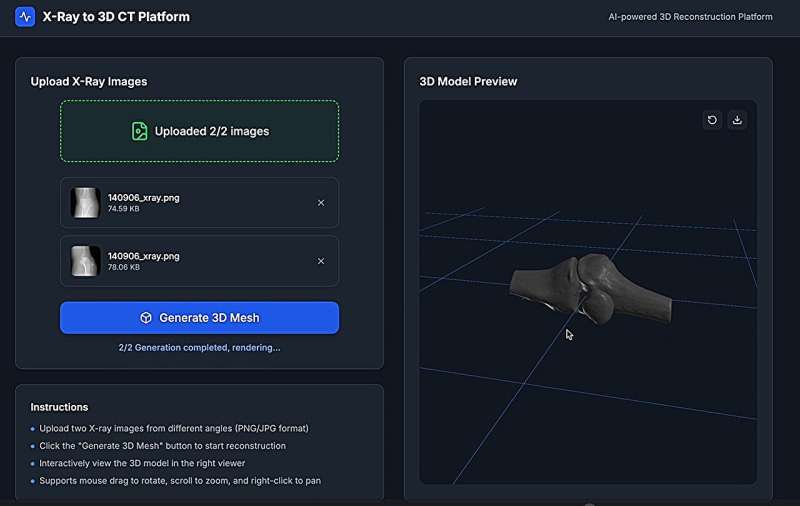
The research team led by Prof. Li Xiaomeng has developed a novel AI-powered algorithm to generate high-quality 3D images of bones and anatomy in under a minute, using as few as 2–4 X-ray snapshots. Credit: Hong Kong University of Science and Technology
Researchers at The Hong Kong University of Science and Technology (HKUST) have developed a groundbreaking AI technology that reconstructs precise 3D bones and organs models from minimal X-ray images, slashing patients' radiation exposure by up to 99% compared to CT scans.
This innovation not only enhances patient safety but also reduces costs and wait times, with potential applications in surgical planning, real-time imaging, and personalized implants. HKUST team is collaborating with different industry partners, including bone printing company Koln 3D Technology (Medical) Limited (Koln 3D), to implement this technology in public hospitals.
Computed tomography (CT) scans are a common medical imaging tool used to diagnose a wide range of conditions, guide surgical procedures, and produce 3D orthopedic and anatomical models to address complex conditions such as deformities, fractures and tumors. However, CT scans expose patients to high levels of radiation, which can be particularly harmful to children, pregnant women and elderly patients who require frequent monitoring.
The HKUST research team led by Prof. Li Xiaomeng, Assistant Professor from the Department of Electronic and Computer Engineering and Associate Director of Center for Medical Imaging & Analysis, has developed a novel AI-powered algorithm that generates 3D bone and anatomical images with less than one hundredth of the X-ray views required for CT scans—drastically cutting down radiation exposure by up to 99%. Key advantages of this new imaging technique include:
To create impact of this breakthrough, Prof Li's team is partnering with Koln 3D—Hong Kong's first orthopedic and metal component printing company—to integrate this new technology into pre-surgical planning (custom bone models), personalized implants (tailored to patient anatomy) and real-time surgical navigation.
Prof. Li said that the new technology has diverse applications, with 3D bone printing being merely the first step. "There is tremendous demand for 3D printing solutions in orthopedics, and rapid, high-definition imaging technology could significantly optimize surgical workflows.
"Our technology is already assisting surgeons in preoperative planning, and we aim to expand into creating implantable bone structures while advancing imaging precision of 3D anatomical structures beyond our current 97% accuracy benchmark compared to conventional CT scans, " she said, adding that the team intends to deploy the technology in areas such as real-time surgical navigation, and personalized treatment planning.
Mr. Edmond Yau, Founder and CEO of Koln 3D, said, "HKUST's AI technology solves our biggest challenge: time. Previously, designing a single implant took days of CT scans and manual modeling, not to mention the wait time for CT scan services at public hospitals. Now, we can generate 3D bone models using just two X-ray inputs in less than 30 seconds. This is transformative for patients needing urgent interventions. This collaboration enables us to deliver personalized solutions at scale, cutting costs by 50%–70% while improving outcomes."
Dr. TSE Lung Fung, Specialist in Orthopedics & Traumatology at a private hospital and Medical Consultant to Koln 3D, added, "In my 30 years of practice, I've witnessed patients endure repeated CT scans just to monitor healing. With this technology, we reduce radiation exposure by over 90% while delivering unprecedented accuracy. For children, pregnant women or elderly patients needing frequent checks, it is revolutionary. Now, we can design implants tailored to a patient's unique anatomy in minutes, ensuring a perfect fit and faster recovery."
A public hospital is set to validate the technology in the coming months. If proven effective, it may be adopted in the public medical system by next year at the earliest. The research team is also seeking collaborations with other hospitals to explore applications in cardiac and pulmonary imaging, aiming to redefine standards for safer, faster diagnostics as well as enhance the efficacy of surgical planning and intraoperative care.


Post comments