
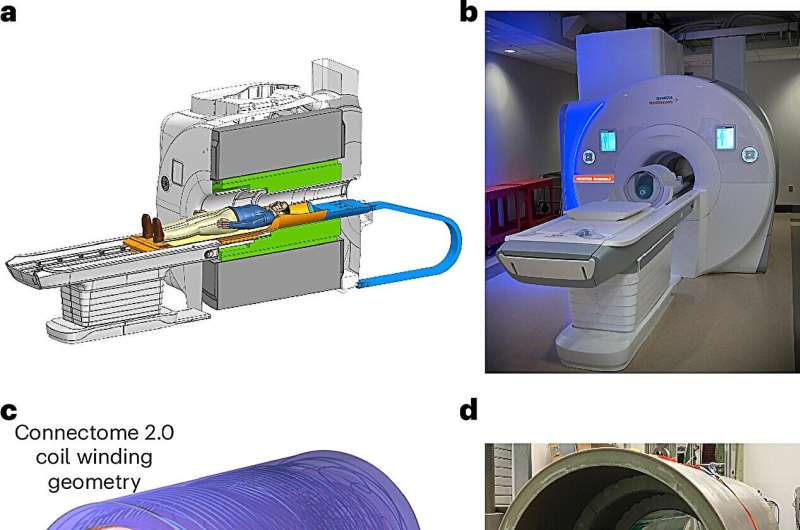
Connectome 2.0 gradient coil. Credit: Nature Biomedical Engineering (2025). DOI: 10.1038/s41551-025-01457-x
A scientific team supported in part by the National Institutes of Health (NIH) has developed a new, ultra-high-resolution brain imaging system that can reconstruct microscopic brain structures that are disrupted in neurological and neuropsychiatric brain disorders. The new system is a significant advance over conventional magnetic resonance imaging (MRI) scanners that cannot visualize these tiny but clinically important structures.
The system, called the Connectome 2.0 human MRI scanner, overcomes a significant hurdle for neuroscientists: being able to bridge different brain regions and probe tiny structures necessary to define the "connectome, " the complex matrix of structural connections between nodes in the nervous system, and to do it noninvasively in living humans. The research is published in Nature Biomedical Engineering.
"This research is a transformative leap in brain imaging—pushing the boundaries of what we can see and understand about the living human brain at a cellular level, " said John Ngai, Ph.D., Director of NIH's Brain Research Through Advancing Innovative Neurotechnologies Initiative, or The BRAIN Initiative.
"The new scanner lays essential groundwork for the BRAIN CONNECTS program's ultimate goal of developing a wiring diagram for the human brain."
The scanner is innovative in two major ways: It fits snugly around the heads of living people, and it has many more channels than typical MRI systems. These advances greatly increase the signal-to-noise ratio of the system, providing much sharper images of very small biological brain structures than previously possible.
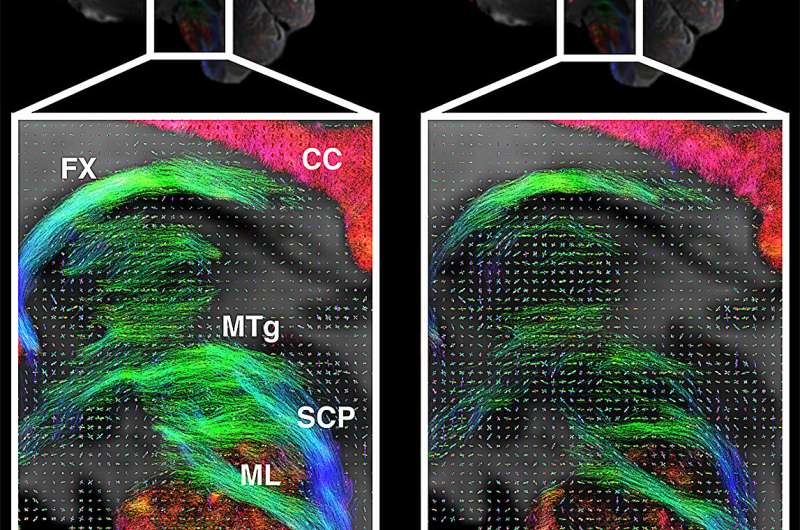
Closeups of the midline sagittal view for Connectome 2.0 (left) and Connectome 1.0 (right) protocols, showing diencephalic and brainstem pathways. Tractography results are shown superimposed onto the underlying fiber orientation distribution functions. Credit: National Institutes of Health
These technical upgrades will enable scientists to map human brain fibers and cellular architecture down to nearly single-micron precision to study how subtle changes in cells and connections relate to cognition, behavior, and disease.
In addition, the team showed that the scanner was safe in healthy research volunteers, revealing subtle microstructural differences (individual axon diameter or cell size) between individual brains. Before this new system, this was only feasible in postmortem or animal studies.
"Our goal was to build an imaging platform that could truly span scales—from cells to circuits, " said senior author Susie Huang, M.D., Ph.D., of the Department of Radiology at Mass General Hospital. "It provides researchers and clinicians with a powerful new tool to study brain architecture in health and disease, in real time."
This work is an important step toward developing a complete wiring diagram of the brain, an achievement that requires novel approaches to map the brain at different scales: across large brain regions and circuits, as well as at the level of tiny cells and connections. It also opens the door for future advances in precision neuroscience, in which noninvasive brain stimulation may help treat brain disorders tailored to an individual's unique brain circuitry.
More information: Gabriel Ramos-Llordén et al, Ultra-high gradient connectomics and microstructure MRI scanner for imaging of human brain circuits across scales, Nature Biomedical Engineering (2025). DOI: 10.1038/s41551-025-01457-x Journal information: Nature Biomedical Engineering


Post comments