
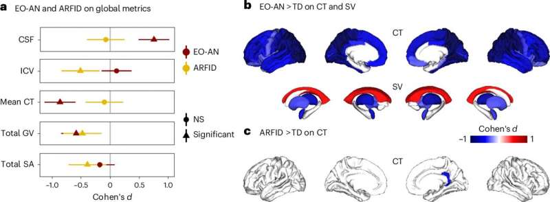
Characterization of the effect of rEO-EDs on brain structure. Credit: Nature Mental Health (2025). DOI: 10.1038/s44220-025-00447-x
In the last decade, the incidence of restrictive eating disorders in children, like anorexia-nervosa and avoidant/restrictive food intake disorders (ARFID), has doubled. These disorders have severe consequences for growing children, resulting in nutritional deficiencies and problems with bone development, statural growth and puberty. Most studies have focused on the effects of these disorders in older individuals, and little is currently known about how restrictive eating disorders affect the brain in children or what mechanisms in the brain might be responsible for this restrictive eating behavior.
To get a better understanding of how these early-onset eating disorders work in the brain, researcher Clara Moreau and her team conducted MRI brain scans on 290 children, of which 124 had been hospitalized for early-onset anorexia-nervosa (EO-AN), 50 had been hospitalized for ARFID, and 116 were children with no eating disorders. All participants were under 13 years old, and those who were hospitalized had very low body mass index (BMI) due to restrictive eating. The results were published in Nature Mental Health.
Although EO-AN and AFRID both result in low BMI and malnutrition due to restrictive eating, they are distinct disorders. EO-AN—as well as later onset anorexia-nervosa—is characterized by restrictive eating arising from a distorted body image, while restrictive eating in AFRID arises from sensory issues, such as a dislike of certain food textures, a lack of interest in food or fear of negative health consequences from food. These differences indicate that the disorders probably arise from different mechanisms in the brain.
Moreau and her team wanted to determine whether the two disorders were characterized by different structural changes in the brain, where they might overlap, and also whether there were similarities with other mental health disorders, like ADHD, obsessive–compulsive disorder (OCD) and autism.
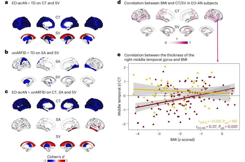
Partition of BMI versus rEO-ED effects on brain features. Credit: Nature Mental Health (2025). DOI: 10.1038/s44220-025-00447-x
After comparing the scans of children with EO-AN and AFRID to those without eating disorders, the researchers found that those with EO-AN showed widespread cortical thinning—thinning of the outer layer of the brain—and increased cerebrospinal fluid compared to those without eating disorders or those with AFRID. They also found a correlation between BMI and cortical thickness in some regions of the brain.
Children with AFRID showed reduced surface area and brain volume and no correlation of changes with BMI. Both AFRID and EO-AN participants had lower gray matter volume than normal. Overall, there were very distinct differences in structural brain changes between the two disorders, despite an initial expectation of overlap between the two.
The team then compared the scans to brain scans of children with ADHD, OCD and autism. They found similarities between those with EO-AN and OCD, and also between those with AFRID and autism. None of the scans showed significant similarities with ADHD data. The authors note this as somewhat surprising, due to inconsistencies with clinical observations showing that around 20% of patients with AN have been found to have autism, and that around 20% of ARFID patients have been found to have ADHD.
This was the first large-scale research study comparing MRI data of these two eating disorders in children. The results have helped highlight the differences between AFRID and anorexia, the mechanisms of these disorders and how they are similar to other mental health disorders. This further understanding of the mechanisms behind these disorders will help to find future treatments or prevention methods.
However, more research is needed to overcome the limitations of this study with regard to the limited AFRID sample size and statistical power. A better understanding of brain restoration after weight normalization would also be helpful for future treatment.
Written for you by our author Krystal Kasal, edited by Gaby Clark, and fact-checked and reviewed by Andrew Zinin—this article is the result of careful human work. We rely on readers like you to keep independent science journalism alive. If this reporting matters to you, please consider a donation (especially monthly). You'll get an ad-free account as a thank-you.
More information: Clara A. Moreau et al, Neuroimaging insights into brain mechanisms of early-onset restrictive eating disorders, Nature Mental Health (2025). DOI: 10.1038/s44220-025-00447-x Journal information: Nature Mental Health
© 2025 Science X Network


Post comments