
by Anne Grimm,Leipzig University
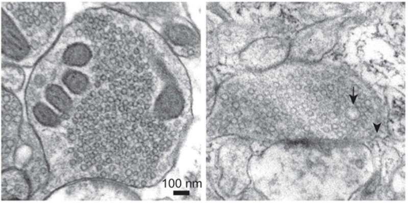
Left: a resting human synapse containing small vesicles that hold neurotransmitters. Right: a synapse that was rapidly frozen 100 milliseconds after an action potential. Arrowhead: membrane invagination forming new vesicles—ultrafast endocytosis. Credit: Chelsy Eddings
Researchers at Leipzig University's Carl Ludwig Institute for Physiology, working in collaboration with Johns Hopkins University, have achieved an important breakthrough in brain research. The so-called zap-and-freeze technique, which allows processes of signal transmission between nerve cells to be visualized within milliseconds, has now been successfully applied for the first time to acute brain slices from both mice and humans.
This will make it possible in the future to investigate how nerve cells adapt their signal transmission while they are active—in other words, how the release of neurotransmitters and the plasticity of nerve cell processes, so-called synapses, change during learning.
Using the zap-and-freeze technique, nerve cells are electrically stimulated and then rapidly frozen just a few milliseconds later. This method makes it possible to capture the movements of cellular components for observation under an electron microscope. In a newly publishedstudyinNeuron, an international research team led by the Carl Ludwig Institute for Physiology at Leipzig University has demonstrated that this new technique also works in intact brain tissue from both mice and humans.
The researchers initially examined brain samples from mice. Using the zap-and-freeze method, they stimulated nerve cells and observed how small vesicles—tiny membrane-bound sacs—are recycled for neuronal communication after releasing neurotransmitters. In healthy brains, synaptic vesicles help transmit information from one cell to another—a process that is crucial for information processing, learning and memory formation.
The researchers then applied the same method to human brain tissue and found that the process occurs in the same way. In both cases, they identified the protein Dynamin1xA at the sites where vesicles are recycled. This shows that the underlying mechanism functions in essentially the same way in mice and humans.
"As a result, we were able for the first time to directly observe how the cell membrane in the human brain is rapidly renewed after neurotransmitters are released. Using this method, we can, in a sense, observe brain cells as they learn. This confirms the high relevance of model organisms for basic research in neuroscience," says Dr. Kristina Lippmann of Leipzig University's Carl Ludwig Institute for Physiology, corresponding author of the study.
Expertise intwo-photon microscopyand electrophysiology at the Carl Ludwig Institute played a crucial role in the study. The researchers in Leipzig adapted the zap-and-freeze method for use with brain slices. Among other findings, Dr. Lippmann and her team discovered that the technique is ideally suited to the targeted stimulation of nerve fibers aligned with the electrical field, such as the parallel fibers in the cerebellum. The researchers also demonstrated that this approach can induce presynaptic short-term plasticity—a key mechanism underlying learning processes in the brain.
In future, the zap-and-freeze technique will be used to investigate the mechanisms of presynaptic short-term plasticity in the cerebellar cortex in greater detail. This region plays a central role inmotor controland provides insights into how the learning brain functions—from development through to age-related and pathological changes.
The current study builds on aprevious publicationinNature Neuroscience, in which the zap-and-freeze method was first tested on cultured nerve cells. The collaboration between Leipzig University and Johns Hopkins University began during a research stay by Dr. Kristina Lippmann in the United States, where she met Professor Shigeki Watanabe, one of the developers of the zap-and-freeze technique.
More information Chelsy R. Eddings et al, Ultrastructural membrane dynamics of mouse and human cortical synapses, Neuron (2025). DOI: 10.1016/j.neuron.2025.10.030 Journal information: Nature Neuroscience , Neuron


Post comments