
Skin cancer pictures can help you spot abnormalities that warrant evaluation
By Angelica Bottaro
Medically reviewed by Doru Paul, MD
Looking at melanoma pictures can help you learn what to look for when you’re doing a skin self-exam at home. Melanoma is the most dangerous (and fatal) type of skin cancer.1 But it is also important to learn how to spot other, more common skin cancers like basal cell and squamous cell carcinomas.
As a general rule, any new lesion should be checked out by a skin specialist called a dermatologist—especially if the lesion is growing or changing. You'll know the difference if you've been doing routine checks yourself.
This article provides pictures of melanoma and other common types of skin cancer. It also discusses skin cancer symptoms and offers some tips on how to do skin cancer self-checks.
Jovanmandic / Getty Images
Melanoma
Melanoma begins in the cells that control the pigment, or color, of your skin.2 It is the most serious form of skin cancer, which can appear as:
Moles
Nodules
Rashes
Scaly patches
Sores that won’t heal
Look for skin growths or patches that are different from other areas of the skin and change over time.3 Early stages of melanoma may look like a new mole, or changes in the shape, size, and color of an existing mole. Sometimes an early sign of melanoma is itching or bleeding of an existing mole.
Melanoma may also appear as a large spot or patch on the skin. Melanomas tend to have irregular borders and may include a range of colors from brown and blue to red, pink, or white.
Most melanomas are flat or slightly raised. Besides the way they look, they don’t usually have symptoms. These pictures of melanomas will help you distinguish between the four main types.
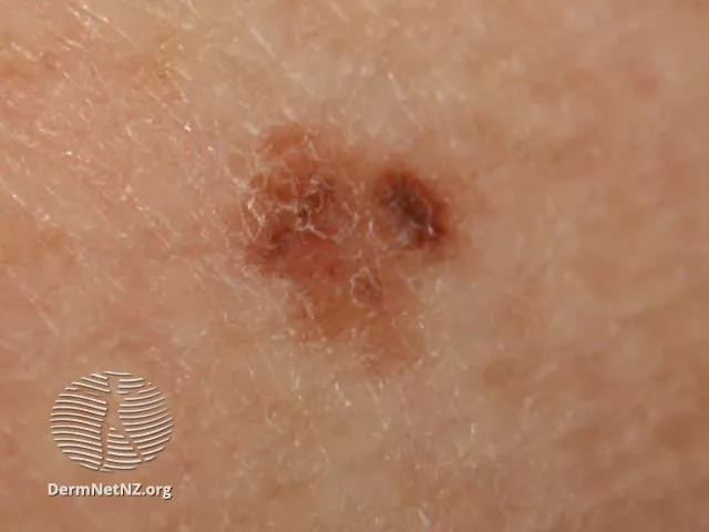
DermNet NZ
Risk factors for melanoma include:
Sun exposure
Fair skin
Family history of melanoma
Some research suggests that genetics play a role in 72% of cases.4
The ABCDE Method to Check for Melanoma
Use the ABCDE method to check for this form of cancer:
Asymmetry: Normal moles tend to be symmetrical, or a similar shape all the way around. If a mole is asymmetrical, it could be a sign of melanoma.
Border: Harmless moles will have regular edges. Those that could be melanoma often have an irregular border.
Color: The color of a mole can be a good indicator of whether it needs to be checked. Melanoma moles will have more pronounced coloring that varies. They can be red, black, dark brown, or flesh-colored.
Diameter: The size of the mole matters. If a mole is larger than the eraser end of a pencil, it should be checked.
Evolving: Moles that change over time may need to be checked. Changes in color, size, shape, or elevation should always be examined by a dermatologist.
The ABCDE method can help you keep track of any mole changes that require a visit to the dermatologist.
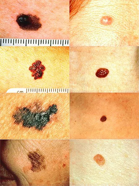
Comparison of melanomas (left column) with normal moles (right column), illustrating parts A, B, C, and D of the "ABCDE" rule (from top to bottom: Asymmetry, Border irregularity, Color variation, and Diameter greater than 6 millimeters). Image courtesy of Wikimedia Commons/Stevenfruitsmaak
Using Ugly Duckling Signs to Check for Melanoma
The “ugly duckling sign” is an observation method that helps people identify a mole that could be cancerous.
This just means you should look for moles that are “ugly ducklings” compared to your other moles. Any mole that stands out as being different in size, shape, or color compared to your other moles should be checked by a dermatologist.
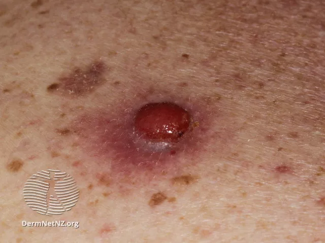
Nodular Melanoma
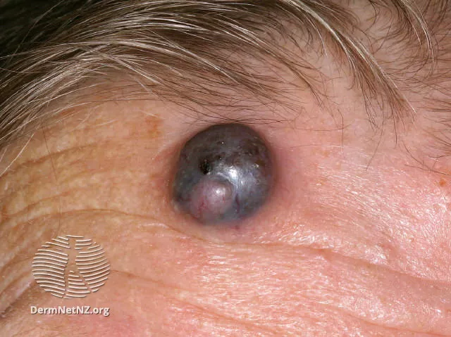
DermNet NZ
Nodular melanoma can develop on any part of the body. However, it occurs most often on:
Legs
Torso
Arms
Head
A nodular melanoma may look like a mole. It can also look like a bug bite, pimple, or blood blister. While a bug bite, pimple, or blood blister will heal over a few days, however, a nodular melanoma does not go away.
Nodular melanomas are typically solid in color. It is often black, but it can also be pink, tan, blue, gray, red, or white.
Assigned males are more likely to develop nodular melanoma than assigned females. The condition is often found in adults over 50.
The EFG method can be used to detect this type of melanoma:
Elevation: A mole that is elevated off the skin could be a cause for concern. The elevation could be even or uneven.
Firm: Nodular melanomas are usually very firm to the touch.
Growth: Mole growth is a significant cause for concern. This always requires further inspection.
Nodular melanomas are fast-growing. A nodular melanoma will continue to grow past the typical two to three week growth of a new, normal mole.
Dysplastic Nevi
Most moles are benign, but about 10% of people in the United States have moles called dysplastic nevi that may carry a greater risk for developing skin cancer. It is sometimes called an "aytpical mole" and is often bigger (more than 5 millimeters across) than a common mole.5
The dysplastic nevus can be pink, dark brown, or a mixture in between. It's typically flat and smooth, with an irregular border edge and a slightly scaly feel.
The risk of melanoma is about 10 times greater for someone with more than five dysplastic nevi than for someone who has none.5
Amelanotic Melanoma
DermNet NZ
An amelanotic melanoma often has little to no color. It may have a pinkish or whitish appearance.
This type accounts for the majority of melanoma cases in children. It may be difficult to spot using the ABCDE method. That’s because this type of melanoma doesn’t have the typical features of other types of melanoma.
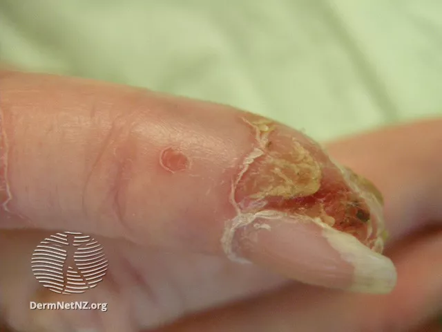
Acral Lentiginous Melanoma
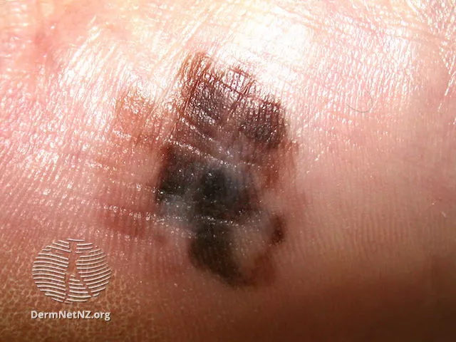
DermNet NZ
Acral lentiginous melanoma occurs on the palms of the hands, soles of the feet, or beneath the nails. It can develop on its own or within an existing mole.
This type of skin cancer first appears as a discolored flat patch. It can infiltrate the skin when it passes from the top layer of skin into the dermis.
This type of melanoma typically looks like a large mole with a smooth surface. It thickens over time. Its color varies from a mixture of brown, blue, and grey to black and red colors.
This cancer is the most common form of skin cancer in people with black or brown skin, accounting for up to 75% of all melanoma cases in this group.6
Assigned males and assigned females are equally affected. The majority of cases occur in adults over the age of 40.
Subungual Melanoma
Subungual melanoma (also called nail apparatus cancer) is uncommon but does occur more in darker-skinned people.7 Beyond genetic factors, though, these cancers also may occur for reasons including injury or trauma to a nail, or as a result of hair salon UV lamp exposure.8
Symptoms include a dark streak (black, blue, or brown line) that runs parallel with the nail, though some may have no color. More lines typically appear over time.
Subungual melanoma often is advanced by the time it is diagnosed, which can contribute to a poor prognosis (outcome). Don't hesitate to tell your healthcare provider as soon as you have concerns.
Basal Cell Carcinoma
Other types of skin cancer look different than the melanoma pictures above. While melanoma is often brown or black in color, other skin cancers are typically pink, red, or skin-colored.
Basal cell carcinoma is the most common type of skin cancer. It develops in the lowest part of the outer layer of skin and is often caused by sun exposure. Basal cell carcinomas can differ in appearance from person to person. Because basal cells grow slowly, with early detection and treatment, most cases are curable.9
Nodular Basal Cell Carcinoma
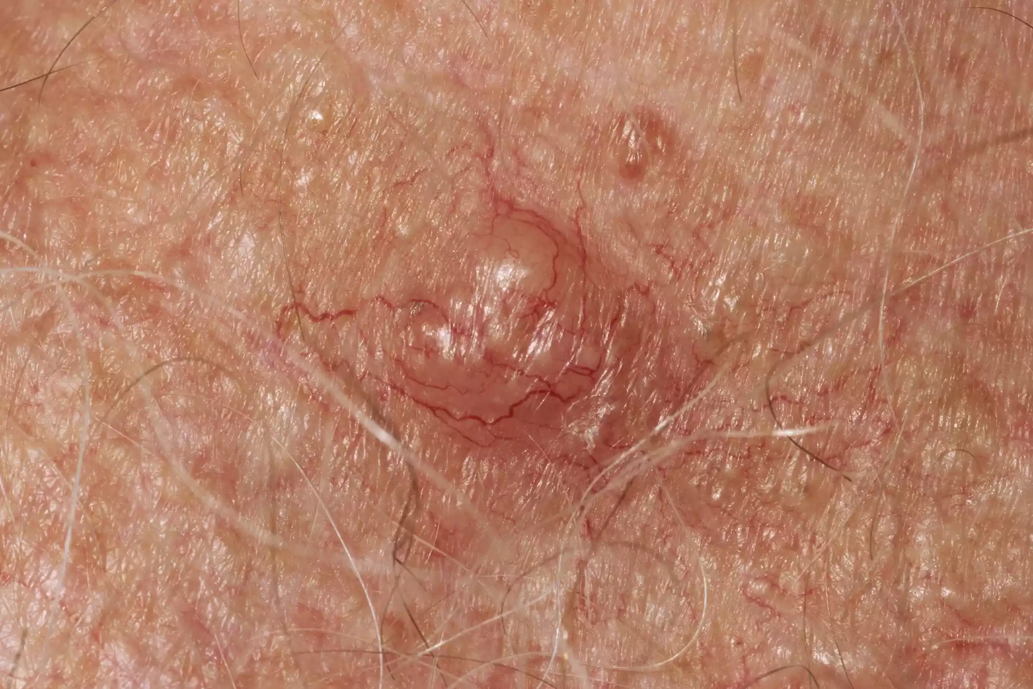
P. Marazzi / Getty Images
Nodular basal cell carcinoma is most often found on the head. This type of cancer starts in basal cells. These cells make new skin cells and push the old ones towards the skin’s surface.
Nodular forms are most common in some nations, including 60% of all cases in the UK, but not in all countries.10 In the United States, it’s estimated that 3.6 million cases of basal cell carcinoma are diagnosed every year. Of those cases, about 2.1 to 2.8 million are nodular basal cell carcinoma.
This type of cancer appears as a pearl-like papule, or swelling. It is round and surrounded by threadlike red lines made up of tiny blood vessels.
Spending a lot of time in the sun increases your risk of developing nodular basal cell carcinoma. Other risk factors include:
Having fair skin
Getting older
Family or personal history of skin cancer
Taking drugs that suppress the immune system
Long term exposure to arsenic, a type of heavy metal
Certain rare genetic disorders such as basal cell nevus syndrome, a condition that impacts the organs throughout the body
Living in high-altitude and sunny locations
Radiation therapy, or treatment that kills cancer cells and shrinks tumors
Although this type of cancer is common, it is highly treatable. The five-year relative survival rate is 100%.11
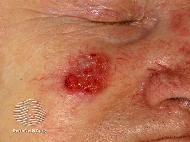
Infiltrative Basal Cell Carcinoma
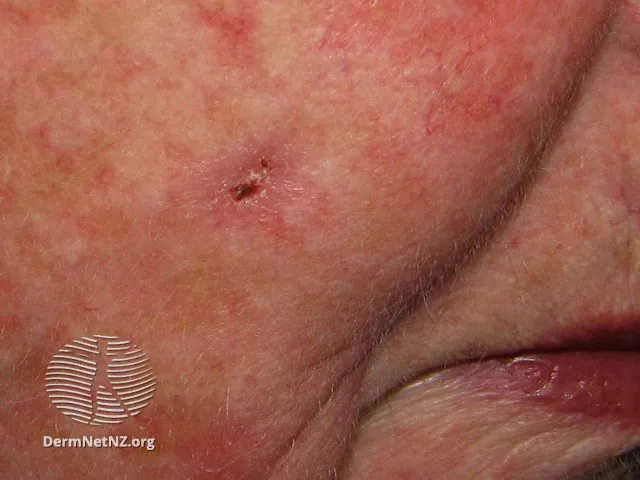
DermNet NZ
Infiltrative basal cell carcinoma occurs when a tumor, or abnormal growth, makes its way into the dermis. The dermis is the inner layer of the two main layers of skin.
Typically, infiltrative basal cell carcinoma appears as scar tissue or thickening of the skin.
Because of its location, this type of skin cancer is harder to diagnose and treat. It is also aggressive, which means it grows and spreads quickly. It requires a biopsy, or tissue or cell sample, to properly diagnose.
A specific type of surgery called Mohs is used to remove this type of basal cell carcinoma. During surgery, thin layers of skin are removed until there is no cancer tissue left.12
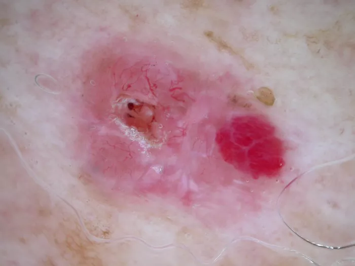
Superficial Basal Cell Carcinoma
DermNet NZ
Superficial basal cell carcinoma is also called in situ basal-cell carcinoma. It is most common on the shoulders or the upper part of the torso. It can also be found on the legs and arms.
Superficial basal cell carcinoma isn’t generally invasive. This means it doesn’t spread to other parts of the body. It grows slowly and is fairly easy to spot and diagnose. It is reddish or pinkish in color and may crust over or ooze.
Superficial basal cell carcinoma accounts for roughly 15%–26% of all basal cell carcinoma cases.13
Squamous Cell Carcinoma
Squamous cell carcinoma occurs when the squamous cells become cancerous. These are small, flat cells in the middle and outer layers of the skin.
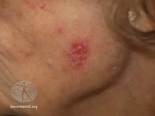
Early Stage Squamous Cell Carcinoma
DermNet NZ
Early stage squamous cell carcinomas may appear as a bump or a flat, scaly patch.
This type of cancer has an extremely high survival rate. It can be aggressive in nature, though. Left untreated, it can spread to other parts of the body and cause serious complications.
Squamous cell carcinoma is mostly found on the parts of the body that get the most exposure to sun, such as:
Face
Lips
Ears
Scalp
Shoulders
Neck
Backs of the hands
Forearms
It can also develop within scars, sores, or skin that has otherwise been damaged in some way.
In the early stages, a nodule will form. The nodule resembles an opalescent wart. This nodule may also have a dip in the middle that looks like a crater.
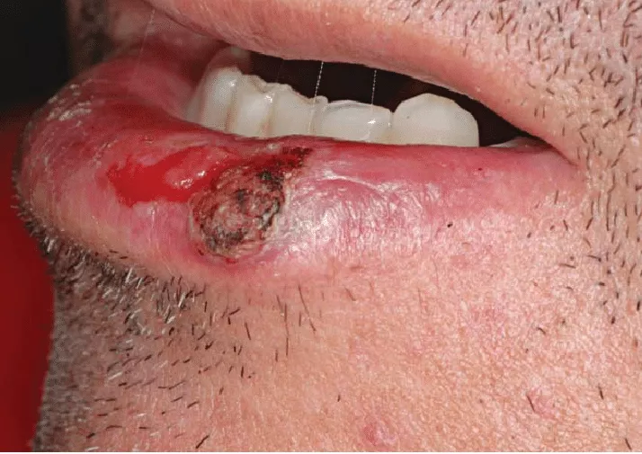
Lip Cancer
Squamous cell carcinoma is the most common type of lip cancer. It starts as a raised, wart-like bump that may or may not have a dimple in the middle.14 A scab-like crust may form, but the area won’t heal and may occasionally bleed.
Squamous Cell Carcinoma With Central Hyperkeratosis
DermNet NZ
Later-stage squamous cell carcinoma has a distinct appearance. The wart-like nodule changes into scaly, red patches called hyperkeratosis. It can also present as an open sore.
When this happens, the crusted skin can bleed on and off and become itchy.
It is important to seek treatment at or before this stage. This is because squamous cell carcinoma may spread in the body. This can lead to more serious health concerns.
Squamous cell cancers affect roughly one million Americans every year. Assigned males are more likely to develop this type of cancer. People over 50 are also at a greater risk.15
Other risk factors include:
Light skin, hair, and eyes
A weakened immune system
Chronic, or long-term infection
Blood cancer
Cancer of the bone marrow, or the spongy tissue within certain bones
Organ transplant, a surgery that replaces a diseased organ with a healthy one
Skin injury or damage
People with xeroderma pigmentosum are also at a greater risk. This is a rare genetic condition that affects the body’s ability to repair DNA, or genetic material, in the skin after sun damage.
Future FamDoc/Wikimedia Commons/CC-BY-SA-4.0
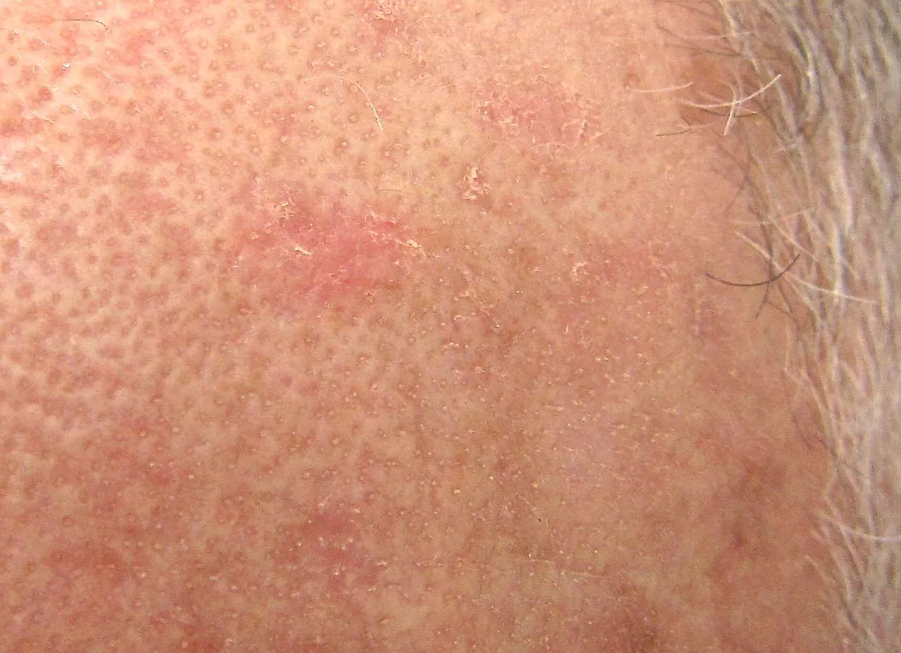
Actinic Keratosis
Actinic keratosis is another type of keratosis that can lead to squamous cell skin cancer. It occurs most common in fair-skinned people, middle-aged or older, with one or more skin lesions.16
The spots feel rough and dry. They appear tan or pink, and often are found on the face, ears, lips, or nose. This is because of damage due to sunlight exposure.
An estimated 58 million people in the U.S. have one or more of these skin lesions, and between 40% and 60% of squamous cell carcinomas arise from these lesions. If untreated, 5% to 10% of these actinic keratosis lesions will progress to cancer.17
Ulcerated Squamous Cell Carcinoma
Research Gate
Squamous cell carcinoma may develop into an ulcer or sore, or become affected by one. This is known as a Marjolin ulcer. Marjolin ulcers can also develop from other cancers, such as basal cell carcinoma, melanoma, or osteosarcoma.18
Marjolin ulcers form in skin that has been damaged in some way. They happen most often in skin that has been badly burned. They can also have other causes, such as:
Bone infections
Pressure sores, also known as bed sores
Frostbite
Skin grafts, a procedure where damaged skin is replaced with healthy skin
Radiation
Marjolin ulcers can take decades to develop. Despite that, this type of cancer is quite aggressive, even if it grows slowly. It can infiltrate, or spread to, other areas of the body.18
In the early stages of this disease, the damaged skin where the ulcer formed will begin to itch and burn. A new sore will show up shortly afterwards.
The new sore is generally flat with hard, raised edges. Other symptoms may occur, such as:
Severe pain
Bleeding
Crusting
Foul-smelling pus
Squamous Cell Carcinoma In Situ
DermNet NZ
Squamous cell carcinoma in situ is also known as Bowen’s disease. It appears as a red or brownish patch or plaque on the skin that grows slowly over time.19 It is a pre-cancerous condition, meaning it could turn into cancer if left untreated.
The patches are often found on the legs and lower parts of the body. They can also be found on the head and neck. In rare cases, the patches are found on the hands and feet, in the genital area, and around the anus.
The condition typically affects Caucasian individuals. Assigned females are more likely to develop Bowen’s disease than assigned males. The majority of cases are in adults over 60.20
As with other skin cancers, Bowen’s disease can develop after long-term exposure to the sun. It can also develop following radiation treatment.
Other causes include:
A suppressed immune system
Skin injury
Inflammatory skin conditions like eczema
A human papillomavirus infection, or a group of conditions that cause warts
Bowen’s disease is generally treatable. It doesn’t usually develop into squamous cell carcinoma. Up to 16% of cases develop into cancer.19
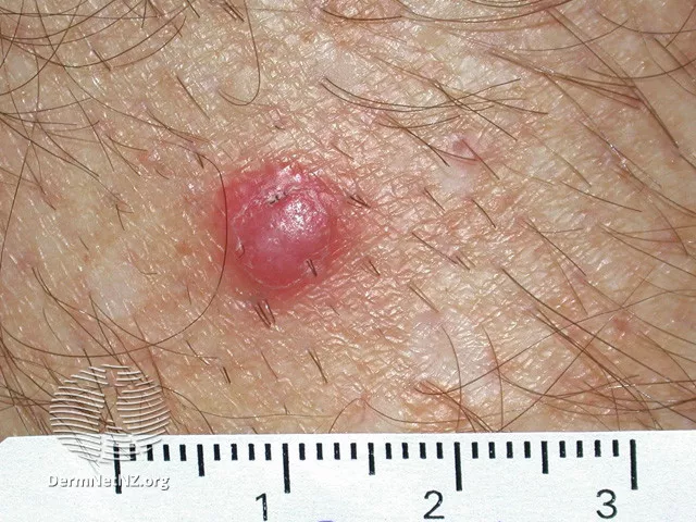
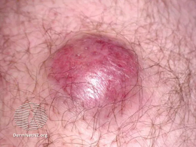
Merkel Cell Carcinoma
Merkel cell carcinoma is rare. It can look similar to the nodular melanoma picture above, but it grows faster.
Merkel cell carcinoma tends to impact the areas of the body that are exposed to the most sunlight. This type of skin cancer presents as a nodule that is flesh-colored or bluish-red.

DermNet NZ
It most commonly found on the face, head, or neck. Older adults are more likely to develop Merkel cell carcinoma.
Risk factors include:
Sun exposure
A suppressed immune system
Light skin
A history of other skin cancers
It is very aggressive and can spread easily throughout the body. Its risk for returning is also high.
It is estimated that one in 130,000 people in the United States will develop Merkel cell carcinoma at some point in their lives.21
DermNet NZ
The AEIOU method can help with early detection:
Asymptomatic: The nodule won’t feel tender.
Expanding rapidly: The nodule grows rapidly in less than three months.
Immunosuppression: A person with a weakened immune system is more at risk for developing Merkel cell carcinoma.
Older age: Adults over 50 are more susceptible to this type of cancer.
UV exposure: The nodule will often appear on sun-exposed, fair skin.
Merkel Cell Carcinoma, Collision Tumor
Europe PMC
A collision tumor occurs when there is more than one type of skin cancer on the same part of the body. This can happen with Merkel cell and squamous cell cancers. It can also happen with Merkel cell and Bowen’s disease or basal cell carcinoma.22
Collision tumors are more likely to occur in adults over 60 following a lifetime of sun exposure.
Other Types of Skin Cancer
Some types of skin cancer are extremely rare. These include:
Lymphoma of the skin, a very rare skin cancer that starts in lymphocytes, white blood cells that help to defend against infections
Kaposi’s sarcoma, a very rare skin cancer associated with human herpesvirus-8 and more likely to occur in people with weakened immune systems2
Why Is Early Detection Important?
The most common cause of skin cancer is prolonged exposure to the sun. However, skin cancer can also develop in areas where there has been no sun exposure.
Early detection of skin cancer is very important and increases the likelihood of survival. This is why knowing the signs and symptoms of skin cancer is so critical. This knowledge can help you decide if you need to get a suspicious looking mole, or skin spot checked out.
Survival rates refer to how likely someone with a specific cancer may live after their diagnosis in comparison to an average person who doesn’t have cancer. Survival rates for skin cancer vary by type:
If melanoma is detected early, individuals are 99% as likely as someone without this cancer to live for at least five years after diagnosis if the cancer is localized and hasn’t spread.23
If basal cell and squamous cell carcinoma are detected early, the survival rate is higher than 95%. (However, these cancers are not reported to cancer registries, so the exact rate is unknown.)24
If Merkel cell carcinoma is detected early, individuals are 76% as likely as someone without this cancer to live for at least five years after diagnosis.25
Tips for Self-Checks
There are steps you can take to prevent skin cancer through routine self-checks. To do a self-check, you should:3
Look at your whole body in a full-length mirror
Check your underarms, forearms, and palms of the hand (nails, too)
Examine your legs, the soles of your feet, and between the toes
You can use a hand mirror to see the back of your head and neck, and your back and buttocks.
You also can use tools like a body mole map to write down your self-exam results, or check out the How to SPOT Skin Cancer™ infographic available from the American Academy of Dermatology. It provides images of the ABCDE features of skin cancers.
Technology also is playing a role in cancer detection, with artificial intelligence (AI) programs now able to identify skin lesions with accuracy comparable to specialists.26 At home, you can take pictures of skin areas to compare any changes over time or share with a healthcare provider.
Summary
Learning how to spot different types of skin cancer can help you get an early diagnosis. Many skin cancers are very treatable in the early stages.
The four types of skin cancer include basal cell carcinoma, squamous cell carcinoma, melanoma, and Merkel cell carcinoma. Appearance may vary from person to person. If you notice nodules, crust, warts, sores, or patches on your body, it’s best to see your dermatologist right away.
Sources
American Academy of Dermatology Association. Skin cancer types: melanoma signs and symptoms.
American Cancer Society. Skin cancer.
American Academy of Dermatology Association. Find skin cancer: How to perform a skin self-exam.
Potrony M, Badenas C, Aguilera P, et al. Update in genetic susceptibility in melanoma. Ann Transl Med. 2015;3(15):210. doi:10.3978/j.issn.2305-5839.2015.08.11
National Cancer Institute. Common moles, dysplastic nevi, and risk of melanoma.
Wang Y, Zhao Y, Ma S. Racial differences in six major subtypes of melanoma: descriptive epidemiology. BMC Cancer. 2016;16(1):691. doi:10.1186/s12885-016-2747-6
Gupta AK, Bharadwaj M, Mehrotra R. Skin cancer concerns in people of color: risk factors and prevention. Asian Pac J Cancer Prev. 2016;17(12):5257-5264. doi:10.22034/APJCP.2016.17.12.5257
LaRocca CJ, Lai L, Nelson RA, Modi B, Crawford B. Subungual melanoma: A single institution experience. Med Sci (Basel). 2021 Sep 15;9(3):57. doi:10.3390/medsci9030057.
Skin Cancer Foundation. Basal cell carcinoma.
Thomson J, Hogan S, Leonardi-Bee J, Williams HC, Bath-Hextall FJ. Interventions for basal cell carcinoma of the skin. Cochrane Database Syst Rev. 2020 Nov 17;11(11):CD003412. doi:10.1002/14651858.CD003412.pub3.
Canadian Cancer Society. Survival statistics for non-melanoma skin cancer.
Chen ELA, Srivastava D, Nijhawan RI. Mohs micrographic surgery: Development, technique, and applications in cutaneous malignancies. Semin Plast Surg. 2018 May;32(2):60-68. doi: 10.1055/s-0038-1642057
Singha J, Patel N. Superficial basal cell carcinoma on the face is a diagnostic challenge. Indian J Dermatol. 2016;61(2):236. doi:10.4103/0019-5154.177802
Skin Cancer Foundation. Ask the expert: What will help me feel less nervous about my lip cancer?
Skin Cancer Foundation. Squamous cell carcinoma risk factors.
Wessely A, Steeb T, Heppt F, Hornung A, Kaufmann M, Koch E, et al. A critical appraisal of evidence- and consensus-based guidelines for actinic keratosis. Curr Oncol. 2021;28(1):950-960. doi:10.3390/curroncol28010093
Skin Cancer Foundation. Actinic keratosis.
Khan K, Schafer C, Wood J. Marjolin ulcer: a comprehensive review. Adv Skin Wound Care. 2020;33(12):629-634. doi:10.1097/01.ASW.0000720252.15291.18
Mohandas P, Lowden M, Varma S. Bowen’s disease. BMJ. 2020;368:m813. doi:10.1136/bmj.m813
Palaniappan V, Karthikeyan K. Bowen's disease. Indian Dermatol Online J. 2022 Mar 3;13(2):177-189. doi:10.4103/idoj.idoj_257_21.
Skin Cancer Foundation. Merkel cell carcinoma.
Hobbs MM, Geers TE, Brown TS, Malone JC. Triple collision tumor comprising Merkel cell carcinoma with an unusual immunophenotype, squamous cell carcinoma in situ, and basal cell carcinoma. J Cutan Pathol. 2020;47(8):764-767. doi:10.1111/cup.13698
American Cancer Society. Survival rates for melanoma skin cancer.
Moffitt Cancer Center. Skin cancer survival rate (nonmelanoma).
American Cancer Society. Survival rates for Merkel cell carcinoma.
Phillips M, Marsden H, Jaffe W, Matin RN, Wali GN, Greenhalgh J, et al. Assessment of accuracy of an artificial intelligence algorithm to detect melanoma in images of skin lesions. JAMA Netw Open. 2019 Oct 2;2(10):e1913436. doi: 10.1001/jamanetworkopen.2019.13436.

By Angelica Bottaro
Angelica Bottaro is a professional freelance writer with over 5 years of experience. She has been educated in both psychology and journalism, and her dual education has given her the research and writing skills needed to deliver sound and engaging content in the health space.


Post comments