
Introduction
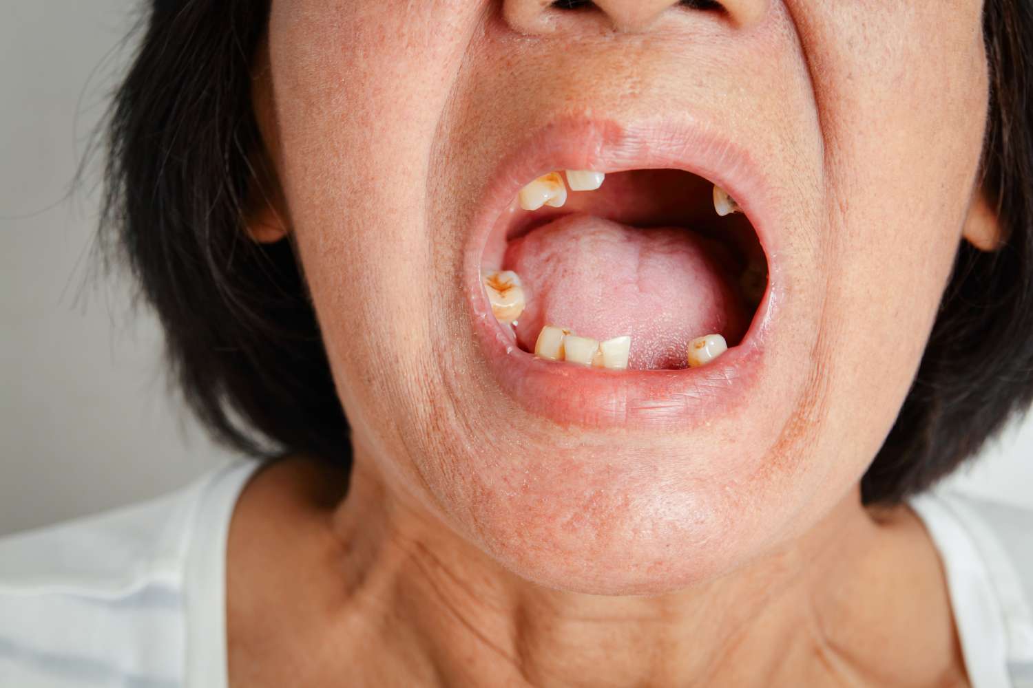
Oral cancer continues to exact a growing toll on global health, with incidence rates rising from 3.26 per 100,000 in 1990 to 5.34 in 2021. This steady increase in both incidence and mortality underscores a critical, persistent gap in early detection and diagnosis. While artificial intelligence (AI) has made remarkable strides in transforming cancer care—enhancing early detection, diagnostic accuracy, and workflow efficiency across diseases like breast, lung, skin, and cervical cancers—oral cancer remains notably underrepresented in this technological revolution.
Despite AI’s promise in medical imaging and digital pathology, its impact in oral oncology has been limited. This blind spot reflects a convergence of complex factors: anatomical and clinical variability, subtle progression of disease, and the diagnostic challenge of differentiating malignant lesions from benign or inflammatory mimics. Compounding these obstacles are the scarcity and heterogeneity of high-quality oral cancer datasets, which hamper the development of reliable, generalizable AI models. Even promising tools like HKU’s OralCancerPredict, which demonstrate encouraging performance in early detection and risk classification, remain geographically limited in accessibility and reproducibility due to disparities in imaging quality, data annotation, and clinical workflows.
If AI is to help close—rather than reinforce—the diagnostic gap in oral cancer, the field must urgently address these multifactorial limitations, from data infrastructure and imaging standardization to clinical integration and equity in access.1,2,3,4
AI’s Promise in Oncology—But Why Not Oral?
Artificial intelligence has already demonstrated tremendous potential in cancer diagnostics, particularly in domains with standardized, high-resolution imaging workflows. In breast cancer, AI-assisted mammography systems now match or exceed double-reader accuracy, with sensitivity and specificity frequently surpassing 90%. These systems not only support accurate detection but also reduce unnecessary recalls and alleviate radiologist workload.
Lung cancer diagnostics have similarly benefited from AI-based CT scan analysis, with meta-analyses reporting area under the curve (AUC) values approaching 0.93—comparable to experienced thoracic radiologists. These tools have enabled more consistent and rapid detection, supporting population-scale screening programs. In dermatology, deep learning algorithms have been shown to enhance clinicians’ ability to identify melanoma and other skin malignancies, especially among non-specialists, thereby expanding diagnostic capacity in primary care and underserved settings.
Cervical cancer screening has also integrated AI, with large-scale studies demonstrating over 89% sensitivity and specificity when AI tools support cytopathologists, leading to earlier detection and improved workflow efficiency.
However, these successes rely on key enablers: structured data, standardized image acquisition protocols, and clearly defined diagnostic workflows. Oral cancer deviates from this model. Its detection is rooted in multimodal clinical assessment—visual inspection, palpation, and ultimately, tissue biopsy. Lesion evaluation depends heavily on clinician intuition and experience, complicating attempts to automate the diagnostic process with image-based AI tools alone. Thus, while AI continues to reshape oncology, oral cancer remains an outlier—an area where automation struggles to replicate the nuanced, tactile, and interpretive elements of expert clinical judgment.5,6,7,8
Why AI Struggles with Oral Cancer
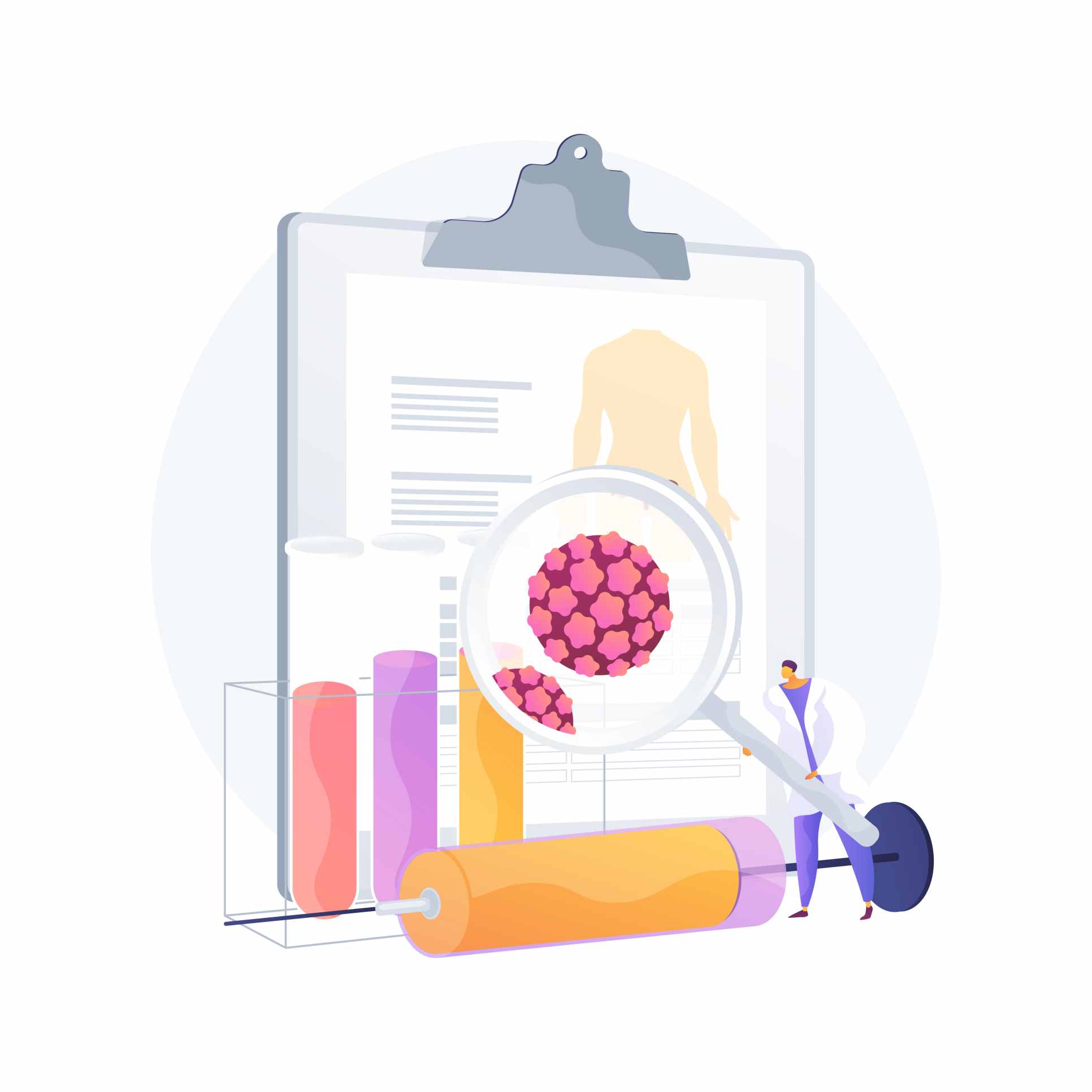
The challenges AI faces in oral cancer detection are multifaceted and deeply rooted in both clinical and technical domains. At the foundation lies a critical shortage of large, annotated, and diverse oral cancer datasets. This limits the ability of AI models to learn from the wide spectrum of lesion types, patient demographics, and imaging variations required for robust generalization.
Unlike the standardized environments in radiology or dermatopathology, oral imaging is often performed under variable lighting conditions, using different equipment, and is prone to motion artifacts and inconsistent focus. The oral cavity’s moist, mobile, and heterogeneous tissue environment adds to this complexity, making consistent image acquisition technically difficult.
Even when high-quality images are obtained, the diagnostic process itself presents unique barriers. Many early oral cancers resemble benign conditions such as traumatic ulcers, candidiasis, or lichen planus. Without reliable cues, AI algorithms trained solely on visual data often struggle to distinguish between malignant and non-malignant lesions. Furthermore, confirmatory biopsies—the clinical gold standard—are not always performed for indeterminate or low-risk lesions, contributing to data labeling uncertainty and model training limitations.
From a research standpoint, oral cancer also receives relatively less attention than other malignancies, particularly in high-income countries. Its burden is disproportionately borne by populations in low-resource settings—those with limited access to research infrastructure, data sharing mechanisms, and technological investment. This structural inequity further restricts the development and validation of AI systems in oral oncology.
Together, these challenges create a feedback loop of underrepresentation, where a lack of data leads to limited research, which in turn delays the creation of effective diagnostic tools. Without strategic intervention to break this cycle—through initiatives focused on standardized data collection, equitable funding, and context-specific AI development—the gap between oral cancer and the rest of AI-augmented oncology is unlikely to close.9,10,11,12
Reference:
Wu J, Chen H, Liu Y, Yang R, An N. The global, regional, and national burden of oral cancer, 1990-2021: a systematic analysis for the Global Burden of Disease Study 2021. J Cancer Res Clin Oncol. 2025 Jan 28;151(2):53. doi: 10.1007/s00432-025-06098-w. PMID: 39875744; PMCID: PMC11775039.
González-Ruiz I, Ramos-García P, Ruiz-Ávila I, González-Moles MÁ. Early Diagnosis of Oral Cancer: A Complex Polyhedral Problem with a Difficult Solution. Cancers (Basel). 2023 Jun 21;15(13):3270. doi: 10.3390/cancers15133270. PMID: 37444379; PMCID: PMC10340032.
Suga T, Tu TTH, Harada H, Toyofuku A. An AI blind spot for detecting oral cancer. Br Dent J. 2025 Jul;239(2):86. doi: 10.1038/s41415-025-9022-7. Epub 2025 Jul 25. PMID: 40715377.
Sapkal R, Supnekar S, Shete A, Prakash Channe P, Desai A. Deep Learning and AI in Detection of Oral Malignancy. J Neonatal Surg [Internet]. 2025Feb.7 [cited 2025Aug.5];14(1S):919-28. Available from: https://www.jneonatalsurg.com/index.php/jns/article/view/1616
Yu SM, Young CYM, Chan YH, Chan YS, Tsoi C, Choi MNY, Chan TH, Leung J, Chu WCW, Hung EHY, Chau HHL. Artificial intelligence-based computer-aided diagnosis for breast cancer detection on digital mammography in Hong Kong. Hong Kong Med J. 2024 Dec;30(6):468-477. doi: 10.12809/hkmj2310920. Epub 2024 Dec 19. PMID: 39697000.
Liu M, Wu J, Wang N, Zhang X, Bai Y, Guo J, Zhang L, Liu S, Tao K. The value of artificial intelligence in the diagnosis of lung cancer: A systematic review and meta-analysis. PLoS One. 2023 Mar 23;18(3):e0273445. doi: 10.1371/journal.pone.0273445. PMID: 36952523; PMCID: PMC10035910.
Krakowski I, Kim J, Cai ZR, Daneshjou R, Lapins J, Eriksson H, Lykou A, Linos E. Human-AI interaction in skin cancer diagnosis: a systematic review and meta-analysis. NPJ Digit Med. 2024 Apr 9;7(1):78. doi: 10.1038/s41746-024-01031-w. PMID: 38594408; PMCID: PMC11004168.
Wang J, Yu Y, Tan Y, Wan H, Zheng N, He Z, Mao L, Ren W, Chen K, Lin Z, He G, Chen Y, Chen R, Xu H, Liu K, Yao Q, Fu S, Song Y, Chen Q, Zuo L, Wei L, Wang J, Ouyang N, Yao H. Artificial intelligence enables precision diagnosis of cervical cytology grades and cervical cancer. Nat Commun. 2024 May 22;15(1):4369. doi: 10.1038/s41467-024-48705-3. PMID: 38778014; PMCID: PMC11111770.
Vinay V, Jodalli P, Chavan MS, Buddhikot CS, Luke AM, Ingafou MSH, Reda R, Pawar AM, Testarelli L. Artificial Intelligence in Oral Cancer: A Comprehensive Scoping Review of Diagnostic and Prognostic Applications. Diagnostics (Basel). 2025 Jan 24;15(3):280. doi: 10.3390/diagnostics15030280. PMID: 39941210; PMCID: PMC11816433.
Chowdhury T, Kasralikar P, Aleem Syed A, Tumati R, Patel S, Kommineni D. Role of Artificial Intelligence in the Diagnosis of Oral Squamous Cell Carcinoma: A Systematic Review. Cureus. 2025 Apr 6;17(4):e81800. doi: 10.7759/cureus.81800. PMID: 40330404; PMCID: PMC12054777.
Singh NN, Tandon A, Jayasankar P. Strength, weakness, opportunities and challenges (SWOC) experience of histopathology image analysis, enhanced by artificial intelligence. J Oral Biol Craniofac Res. 2025 Sep-Oct;15(5):1057-1063. doi: 10.1016/j.jobcr.2025.07.013. Epub 2025 Jul 22. PMID: 40735535; PMCID: PMC12305316.
Veseli, E., Adeoye, J. The future of oral cancer care through AI-powered clinics. Br Dent J238, 908–909 (2025). https://doi.org/10.1038/s41415-025-8902-1


Post comments