
by Haolun Xu, Xi'an jiaotong-Liverpool University
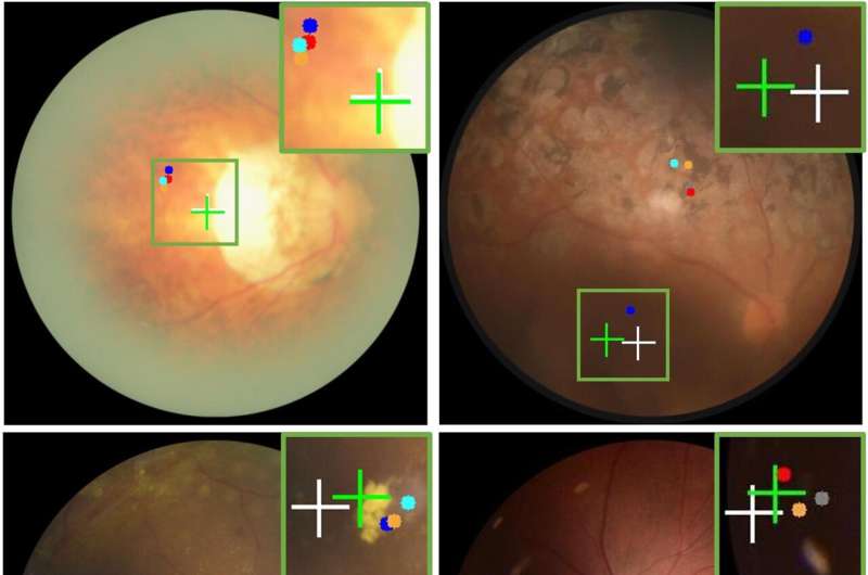
Comparing visual results of fovea localization predictions by various methods. Credit: XJTLU/IEEE 10.1109/JBHI.2024.3445112
Population-based studies have shown retinal disorders are the most common cause of irreversible blindness in developed countries and the second most common cause of blindness after cataracts in developing countries. Medical imaging techniques are key to early detection of retinal disorders, but the technology currently available presents many challenges for practitioners.
To improve the speed and accuracy of diagnoses of retinal disorders, a group of researchers from Xi'an Jiaotong-Liverpool University (XJTLU) and VoxelCloud Inc. in China, has introduced an AI-powered medical imaging technique—DualStreamFoveaNet (DSFN) to address the current imaging challenges. The research is published in the IEEE Journal of Biomedical and Health Informatics.
Dr. Sifan Song, a Ph.D. graduate from XJTLU's School of AI and Advanced Computing and first author of the study, says, "Our new imaging technique, DSFN, has the potential to aid quick and accurate diagnosis of retinal disorders. It also has the potential to be used for other medical conditions that require anatomical structure-based disease diagnosis. For example, in lung cancer screening."
DSFN combines retina images with vascular distribution information to accurately locate the fovea—a depression at the back of the eye where visual acuity is at its highest—in complex clinical scenarios.
Dr. Sifan Song says, "Accurate localization of the fovea allows medical professionals to detect early signs of ocular diseases, such as tiny changes or deposits in the macular region that surrounds the fovea. This helps to regularly monitor disease progression, evaluate the effectiveness of treatment, or adjust treatment plans, and can prevent retinal disorders that lead to irreversible vision loss.
"However, the current medical imaging techniques for identifying fovea location have many limitations."
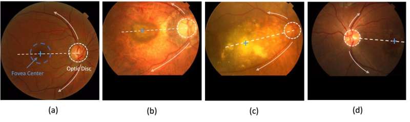
Illustration of the shared global anatomical relationships among the fovea, optic disc, and main blood vessels, in normal (a), diseased (b,c), and poorly conditioned (d) retina images. Credit: XJTLU/IEEE 10.1109/JBHI.2024.3445112
Dr. Song explains that the surrounding retinal tissue's color intensity makes the fovea's dark appearance indistinguishable from the retinal background, which is further obscured by retinal diseases.
He emphasizes that low light conditions and non-standard fovea locations during photography further challenge accurate fovea localization.
"Blurred and poorly lit images make visualizing the back of the eye difficult and may lead to a misdiagnosis. The DSFN helps to overcome many of these challenges," Dr. Song adds.
Dr. Song explains the design of DSFN reduces computational costs while maintaining high accuracy, making it more suitable and affordable for application in clinical environments.
"Lower computational costs are accompanied by faster processing speeds, allowing doctors to obtain diagnostic results more quickly and enabling faster model updates and iterations that lead to more accurate predictions of ocular diseases," says Dr. Song.
Dr. Song is a postdoctoral researcher working at Harvard Medical School and Massachusetts General Hospital.
More information: Sifan Song et al, DualStreamFoveaNet: A Dual Stream Fusion Architecture with Anatomical Awareness for Robust Fovea Localization, IEEE Journal of Biomedical and Health Informatics (2024). DOI: 10.1109/JBHI.2024.3445112.
Provided by Xi'an jiaotong-Liverpool University


Post comments