
Introduction: Why AI-Driven Imaging Matters
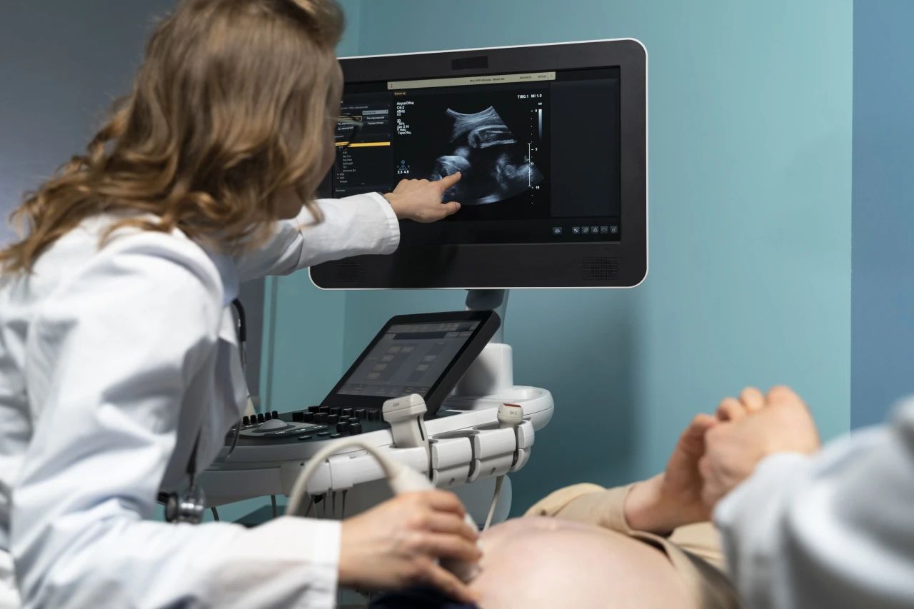
Ultrasound is a cornerstone of diagnostic medicine—safe, portable, and capable of real-time imaging. However, its effectiveness is often constrained by operator dependency, extensive training requirements, and inconsistencies in image acquisition and interpretation.1 These barriers limit its accessibility, especially in low-resource and underserved regions. Artificial intelligence (AI) is rapidly transforming this landscape. By providing real-time guidance, automated quality assessment, and standardized interpretation, AI significantly reduces variability and democratizes access to high-quality imaging.2 Recent studies have shown that even non-expert users can acquire expert-level diagnostic images after brief AI-assisted training. Through deep learning algorithms trained on large datasets, AI systems can identify anatomical landmarks, guide probe positioning, and support lesion detection across organ systems.3 This convergence of AI and ultrasound is redefining scalability, precision, and equity in diagnostic care.
How AI Enhances Ultrasound
While ultrasound has long been valued for its non-invasive, real-time capabilities, it remains susceptible to variability due to differences in operator skill. AI enhances ultrasound by offering intelligent, real-time guidance on probe placement and angle, helping users capture optimal images regardless of experience level.4 Automated interpretation algorithms add another layer of value by identifying conditions such as cardiac dysfunction and fetal anomalies with high sensitivity, thereby supporting faster and more consistent clinical decision-making.5 Deep learning further refines imaging by filtering noise and enhancing contrast, improving overall image quality and diagnostic confidence.6 Notable examples include Butterfly iQ+, which integrates AI for portable, multipurpose scanning, and Caption Health, whose FDA-cleared platform enables guided echocardiographic image acquisition in both inpatient and remote care settings. These advances transform ultrasound into a more consistent, scalable diagnostic tool capable of improving both workflow efficiency and patient outcomes.7
How AI-Guided Ultrasound Works
AI-guided ultrasound systems utilize convolutional neural networks (CNNs) to interpret live image data in real time. These models provide dynamic feedback through visual and textual cues that guide users through probe manipulation, image capture, and anatomical recognition.8 Key features include intelligent probe tracking, automated identification of key structures, auto-capture of optimal frames, and real-time quality scoring to ensure diagnostic accuracy. For instance, Caption Health’s system provides step-by-step instructions for echocardiography, allowing novices to produce expert-level studies.9 Butterfly Network’s AI platform similarly assists in cardiac imaging by predicting probe orientation and suggesting adjustments for better visualization. In addition to acquisition support, CNNs enhance image clarity through denoising and artifact reduction, further improving diagnostic reliability. These capabilities reduce reliance on highly trained personnel and allow ultrasound to be used effectively across diverse clinical environments.10
Clinical Applications and Use Cases
AI-guided ultrasound is increasingly applied across a wide range of clinical settings, from tertiary hospitals to community clinics and field medicine. Its greatest impact is seen in point-of-care scenarios, where rapid, accurate imaging can significantly influence immediate clinical decisions. In emergency care, AI supports quick assessments for conditions like deep vein thrombosis, pericardial effusion, and pneumothorax.11 In primary and rural care, it allows clinicians without radiology expertise to perform diagnostic scans with confidence. In obstetrics, AI enhances fetal anomaly detection and gestational assessments.12 During the COVID-19 pandemic, AI-assisted lung ultrasound provided a crucial, scalable solution for monitoring pulmonary involvement, particularly in resource-constrained areas. Moreover, AI systems improve workflow efficiency by shortening scan times, reducing the need for rescans, and optimizing staff utilization.13 These advances are not just technological milestones—they represent a shift toward more equitable and efficient healthcare delivery.14
Conclusion: The Future of Ultrasound Is Intelligent
AI-guided ultrasound is rapidly reshaping diagnostic imaging by reducing operator dependency, improving image quality, and enabling automated interpretation. It empowers clinicians across all settings to deliver faster, more accurate care—particularly in resource-limited environments where access to trained sonographers is scarce.By streamlining workflows and minimizing variability, AI enhances diagnostic confidence and supports more consistent, equitable care. As models evolve with better data and greater interpretability, AI-powered ultrasound will become an essential tool in precision medicine—enabling earlier, smarter, and more personalized diagnosis worldwide.15,16
Reference:
Exo Imaging. What Are the Pros and Cons of Ultrasound AI? 2025.
Yan L, Li Q, Fu K, Zhou X, Zhang K. Progress in the Application of Artificial Intelligence in Ultrasound-Assisted Medical Diagnosis. Bioengineering (Basel). 2025 Mar 13;12(3):288. doi: 10.3390/bioengineering12030288. PMID: 40150752; PMCID: PMC11939760.
Cai L, Pfob A. Artificial intelligence in abdominal and pelvic ultrasound imaging: current applications. Abdom Radiol (NY). 2025 Apr;50(4):1775-1789. doi: 10.1007/s00261-024-04640-x. Epub 2024 Nov 2. PMID: 39487919; PMCID: PMC11947003.
SmartAlpha Ultrasound AI. Enhancing Ultrasound Imaging with AI.
Caption Health and Heartbeat Health partnership for AI-powered echocardiograms. Cardiovascular Business, 2022.
Abbasian Ardakani A, et al. AdaRes: A deep learning‐based model for ultrasound image denoising. J Clin Ultrasound. 2023.
Sjogren AR, et al. Artificial intelligence in abdominal and pelvic ultrasound imaging. Diagnostics (Basel). 2024.
Victor Mor-Avi. Real-Time Artificial Intelligence–Based Guidance of Echocardiographic Imaging by Novices: Image Quality and Suitability for Diagnostic Interpretation and Quantitative Analysis.
Hareendranathan AR, Chahal BS, Zonoobi D, Sukhdeep D, Jaremko JL. Artificial Intelligence to Automatically Assess Scan Quality in Hip Ultrasound. Indian J Orthop. 2021 Jul 17;55(6):1535-1542. doi: 10.1007/s43465-021-00455-w. PMID: 35003541; PMCID: PMC8688598.
Enhancing Ultrasound Imaging through Convolutional Neural Networks. AHFE Proceedings,2024.
Speranza G, et al. Value of clinical review for AI-guided deep vein thrombosis diagnosis with ultrasound imaging by non-expert operators. Nat Digit Med. 2025 May 5.
Progress in the Application of Artificial Intelligence in Ultrasound Medicine. Front Med (Lausanne). 2025 Mar 13.
Baloescu C, et al. Artificial Intelligence–Guided Lung Ultrasound by Nonexperts. JAMA Cardiol.2025 Mar 1.
Advances in Ultrasound-Guided Surgery and Artificial Intelligence in Musculoskeletal Diseases. Diagnostics. 2024.
Speranza G, et al. Value of clinical review for AI-guided deep vein thrombosis diagnosis with ultrasound imaging by non-expert operators. Nat Digit Med. 2025 May 5; [PMCID: PMC12031111].
Abbasian Ardakani A, et al. Deep learning-based ultrasound image enhancement: improving diagnostic accuracy. J Clin Ultrasound. 2023 Sep; [PMID: 34567890].


Post comments