
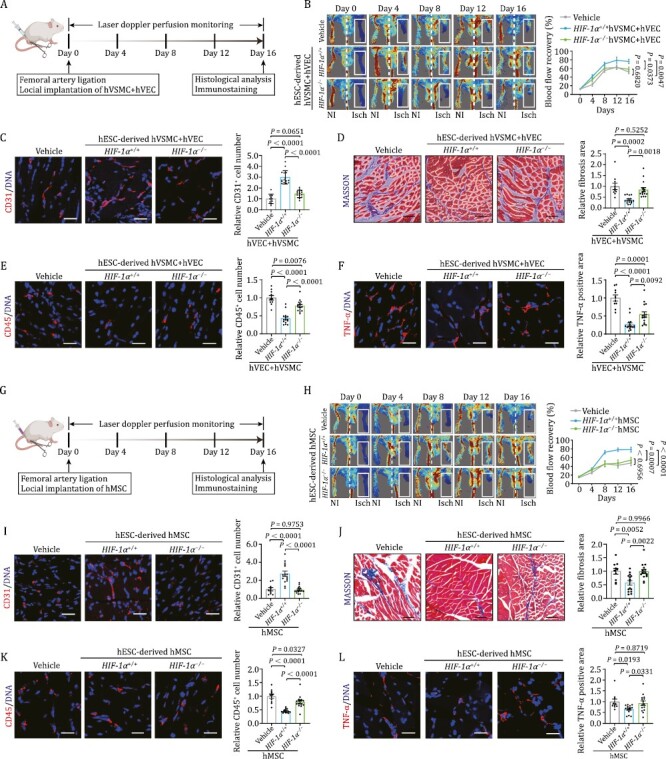
Human vascular cells repair ischemic damage and suppress inflammatory response in a HIF-1α dependent manner. (A) Schematic diagram of the experimental procedures for hVEC and hVSMC transplantation. (B) Representative blood flow images and kinetics of hindlimb ischemic mice injected with vehicle, HIF-1α and HIF-1α+/+−/− cells (hVECs: hVSMCs = 3:1). Laser doppler blood perfusion was measured every 4 days to monitor hindlimb blood flow changes. Data are presented as mean ± SEM. n=10 mice for vehicle group, n = 15 mice for the other groups. Two-way ANOVA followed by Sidak’s test. (C) Representative images of CD31 immunostaining in ischemic hindlimb sections after implantation of HIF-1α or HIF-1α+/+−/− cells (hVECs: hVSMCs = 3:1). Scale bars, 25 μm. Quantitative data are shown as the mean ± SEM. n = 10 mice for vehicle group, n = 15 mice for the other groups. One-way ANOVA followed by Tukey’s test. (D) Representative images of Masson’s trichrome staining in ischemic hindlimb sections after implantation of HIF-1α or HIF-1α+/+−/− cells (hVECs: hVSMCs = 3:1). Scale bars, 25 μm. Quantitative data are shown as the mean ± SEM. n=10 mice for vehicle group, n = 15 mice for the other groups. One-way ANOVA followed by Tukey’s test. (E) Representative images of immunostaining of CD45 in ischemic hindlimb sections after implantation of HIF-1α or HIF-1α+/+−/− cells (hVECs: hVSMCs = 3:1). Scale bars, 25 μm. Quantitative data are shown as the mean ± SEM. n = 10 mice for vehicle group, n = 15 mice for the other groups. One-way ANOVA followed by Tukey’s test. (F) Representative images of immunostaining of TNF-α in ischemic hindlimb sections after delivery of HIF-1α or HIF-1α+/+−/− cells (hVECs: hVSMCs = 3:1). Scale bars, 25 μm. Quantitative data are shown as the mean ± SEM. n = 10 mice for vehicle group, n = 15 mice for the other groups. One-way ANOVA followed by Tukey’s test. (G) Schematic diagram of the experimental procedures for hMSC transplantation. (H) Representative blood flow images and kinetics of hindlimb ischemic mice injected with vehicle, HIF-1α hMSCs and HIF-1α+/+−/− hMSCs. Laser doppler blood perfusion was measured every 4 days to monitor hindlimb blood flow changes. Data are presented as mean ± SEM. n = 10 mice for vehicle group, n = 15 mice for the other groups. Two-way ANOVA followed by Sidak’s test. (I) Representative images of immunostaining of CD31 in ischemic hindlimb sections after implantation of HIF-1α or HIF-1α+/+−/− hMSCs. Scale bars, 25 μm. Quantitative data are shown as the mean ± SEM. n = 10 mice for vehicle group; n = 15 mice for the other groups. One-way ANOVA followed by Tukey’s test. (J) Representative images of Masson’s trichrome staining in ischemic hindlimb sections after implantation of HIF-1α or HIF-1α+/+−/− hMSCs. Scale bars, 25 μm. Quantitative data are shown as the mean ± SEM. n = 10 for vehicle group; n = 15 for the other groups. One-way ANOVA followed by Tukey’s test. (K) Representative images of immunostaining of CD45 in ischemic hindlimb sections after implantation of HIF-1α or HIF-1α+/+−/− hMSCs. Scale bars, 25 μm. Quantitative data are shown as the mean ± SEM. n = 10 mice for vehicle group; n = 15 mice for the other groups. One-way ANOVA followed by Tukey’s test. (L) Re presentative images of immunostaining of TNF-α in ischemic hindlimb sections after implantation of HIF-1α or HIF-1α+/+−/− hMSCs. Scale bars, 25 μm. Quantitative data are shown as the mean ± SEM. n = 10 mice for vehicle group; n = 15 mice for the other groups. One-way ANOVA followed by Tukey’s test.
Ischemic conditions, characterized by reduced blood flow and oxygen supply, pose significant challenges to tissue health and regeneration. Current therapies, including thrombolytics and surgical interventions, often fall short in promoting effective angiogenesis and vascular remodeling. Understanding the mechanisms underlying ischemic repair is crucial for developing novel therapeutic strategies. This review will delve into two recent studies that explore the role of the HIF-1α transcription factor in promoting vascular regeneration and bone repair in ischemic conditions.
Lei et al. investigated the impact of HIF-1α, a core transcription factor responding to changes in cellular oxygen levels, on human vascular cell types and their role in ischemic vascular regeneration. Researchers employed CRISPR/Cas9-mediated gene editing to generate HIF-1α-deficient human embryonic stem cells (hESCs) and directed their differentiation into three major vascular cell types: vascular endothelial cells (VECs), vascular smooth muscle cells (VSMCs), and mesenchymal stem cells (MSCs).
The study revealed that HIF-1α deficiency in all three cell types impaired their ability to promote angiogenesis, indicated by reduced cell migration, tube formation, and capillary density in ischemic tissue. Among the three cell types, MSCs exhibited the highest sensitivity to HIF-1α deficiency, with the greatest changes in gene expression and function under hypoxic conditions.
In addition, the study identified ANKZF1, an effector of HIF-1α, as a key downstream gene that mediated the pro-angiogenic functions of HIF-1α in MSCs. Knockdown of ANKZF1 mimicked the impaired angiogenesis observed in HIF-1α-deficient MSCs.
These findings highlighted the crucial role of HIF-1α in orchestrating the complex interplay between different vascular cell types and their contribution to ischemic vascular regeneration. Understanding the downstream pathways and genes regulated by HIF-1α, such as ANKZF1, may open avenues for developing targeted therapeutic interventions to enhance vascular regeneration in ischemic diseases.
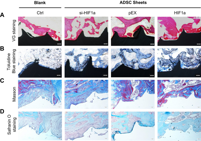
Osteogenesis and new bone formation were observed 30 days after surgery. A VG staining, B toluidine blue staining, C Masson staining, and (D) Safranin O staining under light microscopy. Scale bar = 100 μm.
HIF-1α has also emerged as a promising regulator of osteogenesis and angiogenesis, offering new avenues for enhancing bone regeneration. Song et al. explored the role of HIF-1α in enhancing the osteogenic potential of adipose-derived stem cells (ADSCs) and promoting bone regeneration. Researchers investigated the effects of HIF-1α overexpression and silencing in ADSCs on their proliferation, migration, adhesion, and osteogenic differentiation. Additionally, they evaluated the impact of HIF-1α-modified ADSC sheets on osseointegration and bone formation around titanium implants in a rat model. The study demonstrated overexpression of HIF-1α in ADSCs increased their proliferation, migration, adhesion, and osteogenic differentiation. This was accompanied by upregulation of osteogenic marker genes and proteins. Furthermore, HIF-1α overexpression in human umbilical vein endothelial cells (HUVECs) enhanced their angiogenic potential, indicated by increased tube formation and branching.
In a rat model, HIF-1α-modified ADSC sheets significantly increased osseointegration and bone formation around titanium implants compared to control groups. This was associated with enhanced bone density, thickness, and mineralization rate. Mechanically, the study confirmed that the HIF-1α/VEGF/AKT/mTOR signaling pathway played a crucial role in mediating the enhanced osteogenic potential of HIF-1α-modified ADSCs. HIF-1α upregulated VEGF expression, which in turn activated the VEGF/AKT/mTOR signaling pathway, promoting cell proliferation, migration, and osteogenic differentiation.
These findings provided compelling evidence that HIF-1α played a critical role in enhancing the osteogenic potential of ADSCs by coupling angiogenesis and osteogenesis. The HIF-1α/VEGF/AKT/mTOR signaling pathway serves as a potential target for developing novel therapeutic strategies to promote bone regeneration and improve osseointegration in clinical settings.
The insights gained from these studies offer several potential therapeutic avenues for enhancing vascular and bone regeneration in ischemic diseases:
Targeting HIF-1α for promoting angiogenesis: Developing drugs or therapies that specifically enhance HIF-1α expression or activity could promote angiogenesis and improve blood flow in ischemic tissues. This could have broad applications in treating various ischemic conditions, including cardiovascular diseases, peripheral arterial disease, and stroke.
Modulating the HIF-1α/VEGF/AKT/mTOR signaling pathway for enhancing osteogenesis: Further exploration of the downstream targets and mechanisms of the HIF-1α/VEGF/AKT/mTOR signaling pathway could lead to the development of novel therapeutic strategies for enhancing osteogenesis. This could have significant implications for bone repair and regeneration, particularly in the context of fractures, osteoporosis, and bone defects.
Combining HIF-1α activation with stem cell-based therapies: The use of HIF-1α-modified stem cell therapies, such as HIF-1α-overexpressing ADSC sheets, could enhance the regenerative potential of these cells and improve outcomes in bone repair and regeneration applications.
In summary, both studies underscore the importance of the HIF-1α transcription factor in mediating vascular and bone regeneration responses to ischemic conditions. HIF-1α's ability to regulate gene expression and activate downstream signaling pathways, such as the VEGF/AKT/mTOR pathway, plays a central role in orchestrating the complex cellular processes involved in these regenerative responses. Understanding the intricate mechanisms underlying ischemic vascular and bone regeneration is crucial for developing effective therapeutic strategies.
Reference:
Lei J, Jiang X, Huang D, Jing Y, Yang S, Geng L, Yan Y, Zheng F, Cheng F, Zhang W, Belmonte JCI, Liu GH, Wang S, Qu J. Human ESC-derived vascular cells promote vascular regeneration in a HIF-1α dependent manner. Protein Cell. 2024 Jan 3;15(1):36-51.
Song S, Zhang G, Chen X, Zheng J, Liu X, Wang Y, Chen Z, Wang Y, Song Y, Zhou Q. HIF-1α increases the osteogenic capacity of ADSCs by coupling angiogenesis and osteogenesis via the HIF-1α/VEGF/AKT/mTOR signaling pathway. J Nanobiotechnology. 2023 Aug 7;21(1):257.


Post comments