
By Richard N. Fogoros, MD
Medically reviewed by Jenny Sweigard, MD
When a blood vessel is damaged, your body's blood clotting ability prevents too much blood from being lost. Forming a clot is also the first step in healing the injury.
Sometimes, a blood clot forms when it doesn't need to and blocks a blood vessel. This stops blood from getting to the organs attached to the vessel. When their blood supply is cut off, the organs get damaged and stop working.
The symptoms of organ damage can be the first clue that someone has a blood clot. In some cases, a blood clot can be life-threatening rather than life-saving. That's why it's very important to find out if you have one.
In this article, you will learn about how healthcare providers can diagnose blood clots.
Types of Blood Clots
There are two types of blood clots that can cause serious health problems: a thrombus and an embolus.
A thrombus is a blood clot that forms in a blood vessel. An embolus is a blood clot that travels through a blood vessel and causes a blockage somewhere else in the body.
Blood clots damage tissue because they block blood from flowing through vessels. When the tissue doesn't get oxygen and nutrients from blood, it can lead to conditions like a stroke.
A thrombus or embolus can lead to several health conditions:1
A stroke often happens because there is a thrombus of an artery that goes to the brain. It can also happen if an embolus travels to the brain. These clots often start in the heart or an artery in the neck (carotid artery).
A heart attack is often caused by a thrombus that forms in a heart artery. It's usually made up of substances called atherosclerotic plaque.
A deep vein thrombosis (DVT) is a clot that forms in one of the major veins of the leg, thigh, or pelvis. A DVT can also form in some of the vessels in the arms or upper extremities.
A pulmonary embolus is a blood clot that travels to the lungs. It usually starts as a DVT.
A thrombus in a major vein that drains the liver (portal vein thrombosis).
A thrombus in a vein that drains a kidney (renal vein thrombosis).23
The treatment for a blood clot depends on where it is. One of the most common treatments is a type of medication called blood thinners.
These medications are good at breaking up clots or keeping them from getting bigger. However, they also have serious side effects. People who take blood thinners are at risk for bleeding problems.
Recap
The health problems caused by blood clots depend on where they form. When the blood supply to an organ is blocked by a clot, it damages the organ. Blood clots that form in one place can also move through the bloodstream and cause a blockage somewhere else.
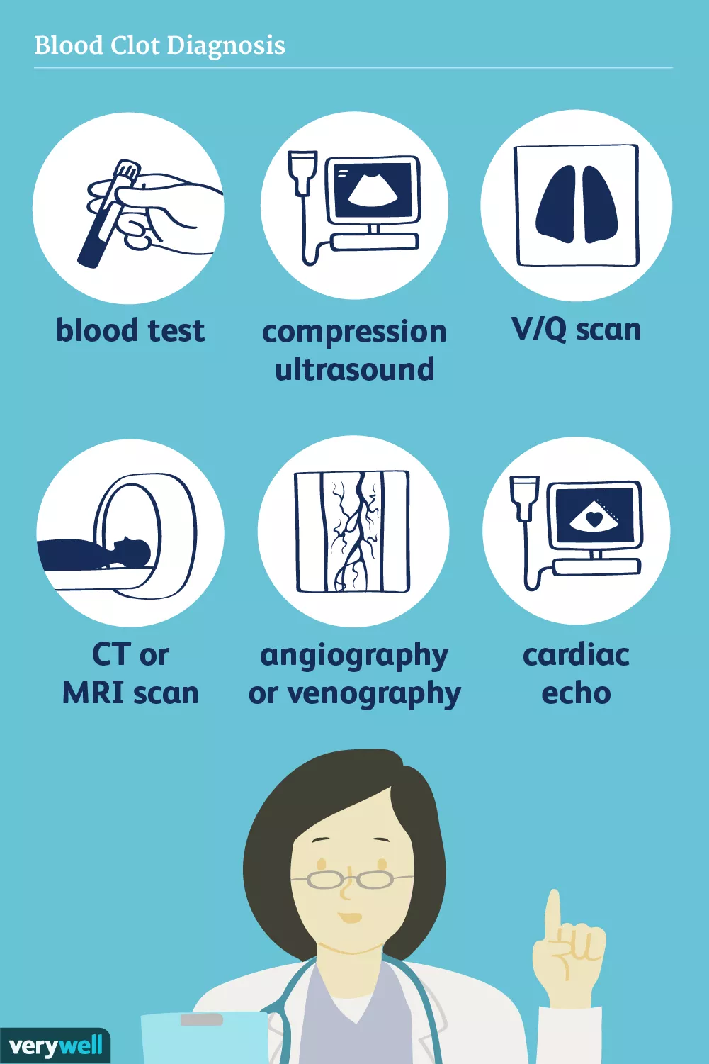
Lab Tests
There are a few lab tests that healthcare providers can use to diagnose a blot clot. The tests can tell if your body's blood clotting system isn't working right.
D-Dimer Blood Test
A D-dimer blood test can tell if there has recently been abnormal clotting activity somewhere in the bloodstream. The test can help providers pin down a diagnosis of a DVT or pulmonary embolus.4 However, the D-dimer value is not specific to a blood clot. It is possible to have an elevated D-Dimer but not have a blood clot.
Cardiac Biomarkers
Cardiac biomarkers help diagnose a heart attack. While the blood tests do not specifically diagnose blood clots, they can tell if the heart muscle is damaged. That damage could be caused by a plaque in the coronary artery that has become a thrombus and broken free.5
Recap
Blood tests can show if there is abnormal blood clotting activity in the body. They can also show if the heart has been damaged by a clot.
Imaging Tests
Imaging tests can help spot a blood clot. Some of the tests can see if there is damage from a blood clot. There are also tests that can tell whether you are at risk of getting a blood clot.
Compression Ultrasound
A compression ultrasound test is useful for diagnosing a DVT.6 The test is non-invasive, which means there are no needles or cuts. The ultrasound wand goes over the spot on your body that your provider wants to get a picture of.
V/Q Scan
During a ventilation-perfusion scan (V/Q scan), a special dye is injected into the bloodstream. It makes it easier to see how blood is flowing to and from the lungs. The test can be used to see if a pulmonary blood vessel is being blocked by an embolus.7
CT Scan
A computerized tomography (CT) scan is a type of X-ray. It gives provides very detailed pictures of the inside of the body.
For example, a CT scan can help providers see if the brain has been damaged by a stroke. It can also look at the lungs to see if they have been damaged by a pulmonary embolus (PE).8
MRI Scan
MRI scans are helpful for seeing whether there is early organ damage after a person has a stroke, a PE, portal vein thrombosis, or renal vein thrombosis.
However, an MRI takes longer than other imaging tests. If a person is in need of immediate care, a provider might use a CT scan instead.7
Recap
Imaging tests can help find a blood clot. They can also show organ damage from a clot.
Procedures
There are also some other tests providers can use to look for blood clots. These tests use imaging but also have some extra steps.
Angiography or Venography
Angiography and venography are imaging procedures that use special dyes to help "light up" the inside of the body. The dyes are injected into the blood vessel that a provider thinks has a clot in it. Then, imaging machines are used to take pictures of the vessel.7
There are also versions of these tests that look for specific types of blood clots:
Pulmonary angiography can be used to diagnose a pulmonary embolus.
Venography is used to diagnose DVTs.
Cardiac angiography looks for clots in the coronary arteries.
Carotid angiography looks for damage or clots in a blood vessel in the neck called the carotid artery.
Echocardiography
Echocardiograms look at the heart and see how it is working. Providers can also use them to see if a thrombus has formed in the heart.
Having some heart conditions makes clots more likely to show up in certain places. For example, in people with atrial fibrillation, clots are usually found in the left atrium of the heart. In people with dilated cardiomyopathy, the clots appear in the left ventricle.
Echocardiograms can also show problems that could make it easier for an embolus to go in and out of the heart. One example of this type of condition is a birth defect called patent foramen ovale.
The test can also be used to figure out if someone is at risk of having a stroke caused by an embolus.9
Recap
Some tests use dye to "light up" the inside of the body. The pictures from the scans show how blood is moving through the body. They can also spot problems with organs that could make it easier for a blot clot to form or move.
Summary
A blood clot can be a life-threatening medical condition. Sometimes, people don't know they have a blood clot. However, people often have symptoms because clots damage organs and tissues.
If a healthcare provider thinks someone has a blood clot, they can do different tests to make the diagnosis. Lab tests and imaging tests can also help providers figure out the kind of clot a person has.
Sources
Centers for Disease Control and Prevention. Venous Thromboembolism (Blood Clots): What is Venous Thromboembolism?.
Wu M, Schuster M, Tadros M. Update on Management of Portal Vein Thrombosis and the Role of Novel Anticoagulants. J Clin Transl Hepatol. 2019;7(2):154-164. doi:10.14218/JCTH.2018.00057
Mazhar HR, Aeddula NR. Renal Vein Thrombosis. In: StatPearls [Internet]. Treasure Island (FL): StatPearls Publishing.
Weitz JI, Fredenburgh JC, Eikelboom JW. A test in context: D-dimer. J Am Coll Cardiol. 2017;70:2411. doi:10.1016/j.jacc.2017.09.024
Aydin S, Ugur K, Aydin S, Sahin İ, Yardim M. Biomarkers in acute myocardial infarction: Current perspectives. Vasc Health Risk Manag. 2019;15:1-10. doi:10.2147/VHRM.S166157
Gibson NS, Schellong SM, Kheir DY, et al. Safety And Sensitivity Of Two Ultrasound Strategies In Patients With Clinically Suspected Deep Venous Thrombosis: A Prospective Management Study. J Thromb Haemost 2009; 7:2035. doi:10.1111/j.1538-7836.2009.03635.x
Centers for Disease Control and Prevention. Diagnosis and treatment of venous thromboembolism.
Stein PD, Yaekoub AY, Matta F, et al. Resolution Of Pulmonary Embolism On Ct Pulmonary Angiography. Ajr Am J Roentgenol 2010; 194:1263. doi:10.2214/AJR.09.3410
National Heart, Lung, and Blood Institute. Venous Thromboembolism.
National Blood Clot Alliance. Signs and symptoms of blood clots.
Texas Heart Institute. Heart attack warning signs.
Additional Reading
Fields JM, Davis J, Girson L, et al. Transthoracic Echocardiography For Diagnosing Pulmonary Embolism: A Systematic Review And Meta-Analysis. J Am Soc Echocardiogr 2017; 30:714. doi:10.1016/j.echo.2017.03.004

By Richard N. Fogoros, MD
Richard N. Fogoros, MD, is a retired professor of medicine and board-certified in internal medicine, clinical cardiology, and clinical electrophysiology.


Post comments