
Overview
The human hair follicle is an intriguing structure, and much remains to be learned about hair anatomy and its growth. The hair follicle can be divided into 3 regions: the lower segment (bulb and suprabulb), the middle segment (isthmus), and the upper segment (infundibulum). The lower segment extends from the base of the follicle to the insertion of the erector pili muscle (also known as the arrector pili muscle). The middle segment is a short section that extends from the insertion of the erector pili muscle to the entrance of the sebaceous gland duct. The upper segment extends from the entrance of the sebaceous gland duct to the follicular orifice. (See the image below.) [1, 2]
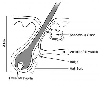
Anatomy of the hair follicle.
The histologic features of the hair follicle change continuously and considerably during the hair growth cycle, thereby making follicular anatomy an even more complex entity.
The size of hair follicles varies considerably during the existence of the follicles. Anagen hairs vary in size from large terminal hairs, such as those on the scalp, to the small vellus hairs that cover almost the entire glabrous skin (except palms and soles). Under hormonal influences, the vellus hair follicles in the male beard area usually thicken and darken at puberty. In predisposed individuals, the terminal hairs on the adult scalp can undergo involutional miniaturization (become vellus).
Although vellus hairs greatly outnumber terminal hairs, the latter are more important. Therefore, the discussion of hair anatomy in this article focuses on terminal hairs.
The follicular life cycle can be divided into 3 phases: anagen, catagen, and telogen. The anagen phase is the phase of active growth, the catagen phase marks follicular regression, and the telogen phase represents a resting period.
In the human scalp, the anagen phase lasts approximately 3-4 years, while the catagen phase lasts about 2-3 weeks, and the telogen phase lasts approximately 3 months. Approximately 84% of scalp hairs are in the anagen phase, 1-2% are in the catagen phase, and 10-15% are in the telogen phase.
Techniques for studying hair microanatomy include the following:
Hair clipping - Performed close to the surface of the scalp
Gentle hair pull
Aggressive hair pluck (trichogram)
Scalp biopsy - Possible use of light microscopy or scanning electron microscopy to study scalp tissue


Post comments