
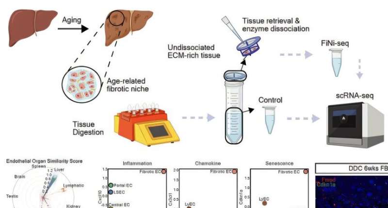
Isolation of fibrotic regions from aged liver tissue, followed by single-cell transcriptome analysis and validation in a fibrosis model. Credit: Nature Aging (2025). DOI: 10.1038/s43587-025-00857-7
Aging and chronic diseases involve the gradual accumulation of subtle tissue changes over a long period. Therefore, there are still limitations in quantitatively understanding these changes within organs and linking them to early signs of disease onset.
In response, Korean researchers have successfully developed a platform technology that accurately captures localized changes that first occur within tissues, significantly aiding in faster disease discovery and prediction, and in setting personalized treatment targets.
A joint research team led by Professor Jong-Eun Park of the Graduate School of Medical Science and Engineering at KAIST and Dr. Chuna Kim of the Aging Convergence Research Center at the Korea Research Institute of Bioscience and Biotechnology (KRIBB) has developed "FiNi-seq (Fibrotic Niche enrichment sequencing)" technology.
The technology captures fibrotic microenvironments locally occurring in aged liver tissue and enables precise analysis at the single-cell transcriptome level.
The research was published in Nature Aging.
The researchers developed a method to selectively enrich early aging microenvironments where regeneration is delayed and fibrosis accumulates, by physically selecting regions with high tissue degradation resistance in aged liver tissue.
In this process, high-resolution identification of fibrosis-related endothelial cells, fibroblasts interacting with the immune system, and immune-exhausted cells such as PD-1 highly expressing CD8 T cells, which were difficult to capture with existing single-cell analysis technologies, was possible.
In particular, the research team confirmed through "FiNi-seq" technology that specific cells observed in fibrotic areas within aged liver tissue secondarily age the surrounding environment through secreted factors, and that this leads to the expansion of the aged environment.
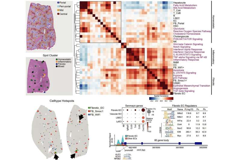
Spatially defined stepwise progression patterns of aging-related regions within the liver and identification of regulatory factors inducing them. Credit: Nature Aging (2025). DOI: 10.1038/s43587-025-00857-7
Furthermore, they also elucidated the mechanism by which endothelial cells lose their tissue-specific identity and induce innate immune responses, promoting immune cell infiltration.
Through spatial transcriptome analysis, the spatial distribution of fibroblasts interacting with immune cells was quantified, revealing their involvement in tissue regeneration, induction of inflammatory responses, and progression to chronic fibrosis.
The research team performed integrated analysis of multi-omics data to obtain transcriptome and epigenome information, precisely interpreting the microenvironment of aged liver tissue and its spatial heterogeneity, and confirming how these changes are connected to the intrahepatic vascular structure.
The newly developed "FiNi-seq" technology is expected to be a useful platform for high-resolution capture of pathophysiological signals in most chronic liver diseases, including the aging process that causes fibrosis.
Dr. Chuna Kim of KRIBB stated, "Through this study, we were able to precisely elucidate the cellular composition and spatial characteristics of the fibrotic microenvironment observed in aged liver tissue at the single-cell level."
Professor Jong-Eun Park of the Graduate School of Medical Science and Engineering said, "As an analytical technology that can capture subtle changes occurring in the early stages of aging and chronic diseases, it is expected to play a significant role in finding effective treatment targets in the future.
"Also, we plan to expand this research to chronic diseases in other organs such as the lungs and kidneys, as well as various liver disease models."
More information: Kwon Yong Tak et al, Quasi-spatial single-cell transcriptome based on physical tissue properties defines early aging associated niche in liver, Nature Aging (2025). DOI: 10.1038/s43587-025-00857-7 Journal information: Nature Aging


Post comments