
Credit:Tumor-derived exosomes drive immunosuppressive macrophages in a pre-metastatic niche through glycolytic dominant metabolic reprogramming.
Non-small cell lung cancer (NSCLC) is a prevalent form of cancer, often linked to the infiltration of immunosuppressive macrophages during metastasis. Exosomes are small extracellular vesicles, ranging from 30 to 150 nanometers in diameter, that can carry various bioactive molecules, including proteins, lipids, and RNA. Tumor-derived exosomes (TDEs) play a crucial role in cancer progression, promoting tumor growth, metastasis, and immune evasion. Cancer cells primarily depend on glycolysis to generate energy and metabolic intermediates, even in the presence of oxygen, a phenomenon known as the Warburg effect. This metabolic reprogramming is not confined to cancer cells but can also occur in immune cells within the tumor microenvironment. Research has shown that tumor cells can induce metabolic reprogramming in macrophages through various mechanisms, causing them to adopt an immunosuppressive phenotype that aids the tumor in evading immune surveillance. However, the mechanisms by which TDEs promote the formation of immunosuppressive macrophages via glycolysis-driven metabolic reprogramming remain unclear. Samantha et al. conducted comprehensive studies using in vitro cell experiments and in vivo mouse models to investigate this mechanism.
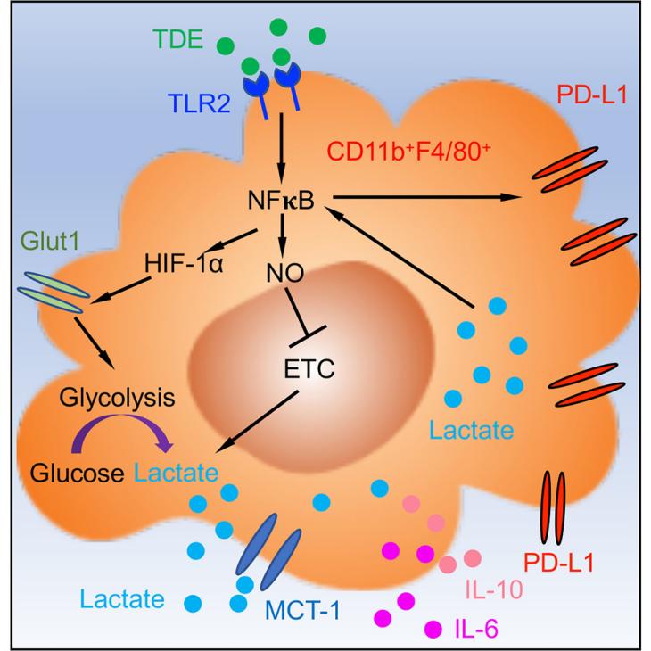
Firstly, researchers isolated exosomes from Lewis lung carcinoma (LLC) cells and treated macrophages with these tumor-derived exosomes (TDEs). Through flow cytometry and immunoblotting assays, they found that TDEs significantly increased the expression of PD-L1 on the surface of macrophages. This mechanism involves the synthesis of PD-L1 driven by the Toll-like receptor 2 (TLR2) and NF-κB signaling pathways, rather than a simple transfer from the exosomes. Additionally, TDEs enhanced glucose uptake in macrophages and upregulated the expression of glycolysis-related genes such as GLUT-1 and LDHA via the NF-κB signaling pathway. Seahorse analysis further validated that macrophages treated with TDEs exhibited higher glycolytic metabolic levels. The study showed that macrophages treated with TDEs produced a large amount of lactate, which, as a metabolic byproduct, activated NF-κB through a feedback mechanism, further enhancing PD-L1 expression. Blocking the lactate channel (MCT-1) significantly reduced PD-L1 expression, indicating that lactate plays a crucial role in this process. Secondly, in mouse model experiments, the researchers found that mice injected with TDEs had a significant increase in micrometastatic foci in the lungs, along with a marked increase in PD-L1 expression in pulmonary macrophages. Using CRISPR/Cas9 technology to knock out the RAB27a gene in 4T1 breast cancer cells, which reduced exosome secretion, resulted in a significant decrease in immunosuppressive phenotype macrophages in the pre-metastatic niche. This highlights the critical role of exosomes in driving this process. Finally, by using 2-DG to inhibit glycolysis and SEITU to inhibit NOS2, the researchers observed the effects on PD-L1 expression. They found that 2-DG significantly reduced PD-L1 expression in macrophages treated with TDEs, while SEITU restored the oxidative phosphorylation level in macrophages and decreased PD-L1 expression.
This study systematically explores, for the first time, the mechanisms by which tumor-derived exosomes (TDEs) drive the formation of immunosuppressive macrophages through metabolic reprogramming. This approach broadens our understanding of the role of exosomes in the tumor microenvironment and immune regulation. Compared to previous methods, the study proposes an NF-κB-dependent metabolic reprogramming model. Instead of examining the effects of TDEs from a single perspective, it integrates multiple aspects—metabolism, immunity, and signaling pathways—providing a comprehensive mechanistic explanation. Moreover, the study validates its hypotheses through a combination of in vitro and in vivo experiments, spanning from the cellular level to animal models, thereby enhancing the reliability and applicability of its conclusions. Importantly, the application of Seahorse analysis technology and metabolic inhibitors offers a deeper understanding of metabolic mechanisms, aiding in the identification of new therapeutic targets.
However, the study primarily focuses on NSCLC and certain breast cancer models, lacking extensive validation across other tumor types. The components and mechanisms of tumor-derived exosomes (TDEs) may vary significantly among different tumor types, necessitating a broader scope of research. Additionally, although the study investigates the role of TDEs, it does not provide a detailed analysis of the specific components—such as proteins, lipids, and RNA—involved in metabolic reprogramming and immune regulation. The complexity and heterogeneity of exosomal components require more precise characterization.
In conclusion, this study provides new perspectives and a significant theoretical foundation for future research in tumor treatment and clinical applications. Further exploration of the specific components of tumor-derived exosomes (TDEs) and their mechanisms, detailed regulation of metabolic reprogramming, feasibility of clinical translation, and individualized treatment strategies will not only enhance the efficacy of cancer treatment but also advance the entire biopharmaceutical industry.
Reference:
Morrissey, Samantha M., et al. "Tumor-derived exosomes drive immunosuppressive macrophages in a pre-metastatic niche through glycolytic dominant metabolic reprogramming." Cell metabolism 33.10 (2021): 2040-2058.


Post comments