




























byAnglia Ruskin UniversityCredit: Tima Miroshnichenko from PexelsA new study has demonstrated for the first time how and why music can reduce distress and agitation for people with advanced deme
byUniversity of HelsinkiThe performance of human observers agrees with the new dim light detection model where performance is fundamentally limited by retinal thresholding nonlinearities along t
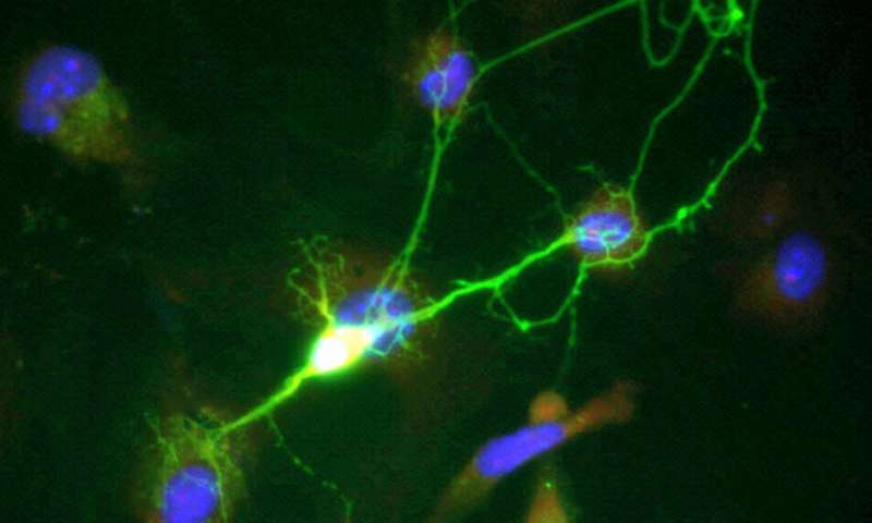
by Society for NeuroscienceA functioning brain cell that is expressing diseased tau. Credit: Hallinan et al.,JNeurosci2019Tau can quickly spread between neurons but is not immediately
byOptica(A) Brightfield photograph of the sample and selected ROI for PS-OCT imaging. (B) PSOCTAverage Intensity measured in dB. (C) PS-OCT Retardance ranging from 0 to π. (D) PS-OCTRetardance A
byUniversity of Technology, Sydney1. Mice are trained to press a left and right lever for pellet and sucrose outcomes (counterbalanced), 2. Mice are prefed to satiety on one of the two outcomes
by University of LeicesterPicture of nerve cells dying in the brain of prion-diseased mice. The nerves are stained pink and can be seen dying in the CA1 region of the hippocampus. Credit: Univers
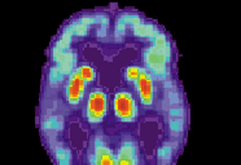
by Scott Lafee,University of California - San DiegoPET scan of a human brain with Alzheimer's disease. Credit: public domainResearchers at University of California San Diego School of
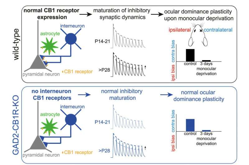
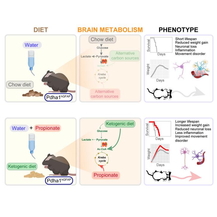
by Netherlands Institute for Neuroscience - KNAWGraphical abstract. Credit:iScience(2024). DOI: 10.1016/j.isci.2024.111410Neuroscientist Rogier Min from the Amsterdam UMC has collaborated