
Scientists identify mutated gene behind mirror movement disorder
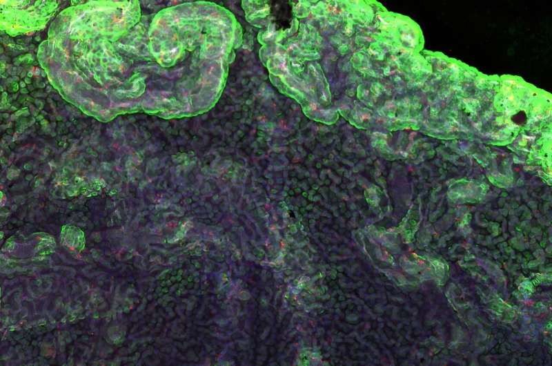
byUniversity of MontrealArhgef7 is required for Netrin-1–mediated commissural axon guidance. (A) The mean mRNA expression, fragments per kilobase of transcript per million mapped reads (fpkm), (
Updated on: April 22,2024
Scientists identify mutated gene behind mirror movement disorder
byUniversity of MontrealArhgef7 is required for Netrin-1–mediated commissural axon guidance. (A) The mean mRNA expression, fragments per kilobase of transcript per million mapped reads (fpkm), (
Updated on:April 22,2024