
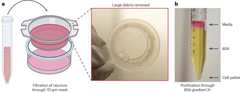
Neuronal purification and generation of a single cell suspension. a, The filtration of neurons through a 70 mm mesh removes large debris from the cell suspension. b, The neuronal purification is achieved by purification through a BSA gradient (2×). Media are placed on top of a 5 mL column of 15% BSA. Following centrifugation, a cell pellet forms at the bottom of the tube. Credit: Nature Protocols (2025). DOI: 10.1038/s41596-025-01194-0
Understanding the electrical activity of neurons is key to unlocking insights into neurological diseases. Yale researchers have unveiled a high-throughput automated method that captures the electrical activity of large numbers of neurons simultaneously and without bias.
This cutting-edge approach provides a powerful "functional fingerprint" of neuron populations in their natural state, opening new doors to understanding and treating neurological diseases. The work was published June 13 in Nature Protocols.
The patch-clamp technique has long been a gold standard for studying the electrical activity of neurons, the fundamental units of the nervous system. However, the manual execution of this approach is slow and labor-intensive. Recent advances in robotic patch-clamp technologies have improved speed and efficiency, but they are limited to artificially grown neurons rather than neurons in their native unmanipulated state.
Developed in the laboratory of Stephen G. Waxman, MD, Ph.D., Bridget M. Flaherty Professor of Neurology at Yale School of Medicine (YSM), the novel methodology offers a streamlined and efficient approach to studying the electrical activity of neurons in their native state while maintaining the accuracy of traditional methods and allowing for large-scale research.
"This method allows us to examine the functional and pharmacological features of neuron populations soon after extraction and to analyze them side by side for direct comparison, " says Mohammad-Reza Ghovanloo, Ph.D., co-lead author of the study, and an associate research scientist in the Department of Neurology at YSM. "By understanding these features, we can make more meaningful comparisons when studying disease mechanisms and gathering clinically useful insights."
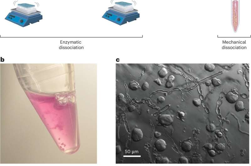
Dissociation of sensory neurons for automated electrophysiology. Credit: Nature Protocols (2025). DOI: 10.1038/s41596-025-01194-0
To complement their innovative approach, the team developed an open-source analysis suite with an intuitive graphical user interface. "This analytical tool enables us to fit data from each neuron with the appropriate biophysical equations, providing a comprehensive functional characterization of the complex, high-dimensional data sets generated by our method, " says Sidharth Tyagi, Ph.D., co-lead author of the study and an MD-Ph.D. student at YSM.
By integrating automation with robust analytical tools, the approach eliminates biases and enhances reproducibility in electrophysiological studies.
The new methodology, which requires six to 18 hours for completion—from preparation of neurons to experimental execution and data analysis—compared to several days with traditional methods, represents a transformative step forward in neuroscience and pharmacology research.
The implications of this breakthrough extend across multiple domains, from fundamental neuroscience to translational applications in drug discovery.
"By enabling high-throughput, simultaneous analysis of large numbers of native neurons, this novel approach paves the way for a deeper understanding of neuronal function and opens the door for the development of targeted therapeutics for neurological disorders, " says Waxman.
More information: Mohammad-Reza Ghovanloo et al, High-throughput multiplex voltage-clamp/current-clamp evaluation of acutely isolated neurons, Nature Protocols (2025). DOI: 10.1038/s41596-025-01194-0 Journal information: Nature Protocols


Post comments