
by Jan Grabowski, Zentrum für Experimentelle und Klinische Infektionsforschung
Sorted microglia exhibit artificially induced transcriptional alterations. Credit: Journal of Neuroinflammation (2024). DOI: 10.1186/s12974-024-03197-2
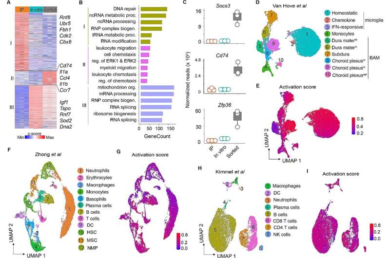
Scientists study the functions of microglia by transcriptomics. This method provides dynamic information about genes that are active in the cell in both health and disease states. To do so, they isolate single cells from tissue followed by an enrichment of selected cell types with fluorescence-activated cell sorting (FACS).
The FACS method is not without its problems though: "We were able to show that the sorted microglia exhibited ex vivo activation signatures," says Dr. Felix Mulenge, a postdoc at the Institute of Experimental Infection Research at TWINCORE and first author of the study, now published in the Journal of Neuroinflammation.
"This is because the cells encounter hydrodynamic stress or traumatic injury during sorting which alters the microglial transcriptome. So we never see the cells in a truly quiescent state."
A long-standing concern is whether such "spurious" signatures are unique to microglia, or whether they are also prevalent in peripheral immune cells. The researchers detected artificial signatures in publicly available data of cells from the CNS as well as other cell types.
They started to wonder whether findings reported in those studies fully reflect the in vivo status of the analyzed cells. "In this era of 'big data,' such ex vivo transcriptional alterations constitute a major technical challenge for many studies," says Mulenge.
To avoid this, Mulenge adopted an alternative method called ribosomal tagging, or RiboTag in short. Instead of sorting the cells to examine the RNA molecules, RiboTag uses a special way of labeling ribosomes of microglia and subsequently enriching tagged ribosomes, which allows determining the translational profile of microglia.
"This strategy requires fewer steps and precludes non-specific microglia activation," says Mulenge. Therefore, the unified set of genes identified here accurately reflect in situ microglia profiles.
"The fact that measurement itself can influence the result of an investigation is a well-known phenomenon in quantum physics," says Prof Ulrich Kalinke, Director of the Institute for Experimental Infection Research and Managing Director of TWINCORE. "With our method, we eliminated the causative sources of artifacts in microglia transcriptomic profiles and created bona fide signatures of microglia."
The researchers hope that their discovery will be used more extensively in the future and thus provide more precise insights into infections in the brain. And not only that: "The method is not limited to the brain in principle, but also works for other peripheral cells," says Mulenge. More informative cell signatures could therefore improve neuroscience as a whole.
More information: Felix Mulenge et al, Transcriptomic analysis unveils bona fide molecular signatures of microglia under conditions of homeostasis and viral encephalitis, Journal of Neuroinflammation (2024). DOI: 10.1186/s12974-024-03197-2
Journal information: Journal of Neuroinflammation
Provided by Zentrum für Experimentelle und Klinische Infektionsforschung


Post comments