
byH. Lee Moffitt Cancer Center & Research Institute
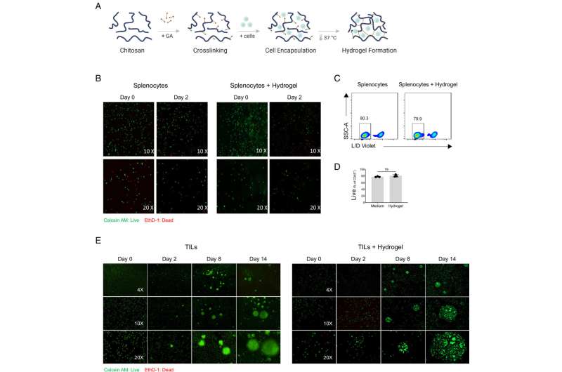
Cell viability assessment in chitosan hydrogel. Schematic representation of chitosan hydrogel preparation and cell and/or MP encapsulation (A). Representative images of mouse splenocytes stained as live (Calcein AM/Green) or dead (EthD/Red) at days 0 and 2 of culture in the complete medium with or without being encapsulated in chitosan hydrogels (B). Representative flow cytometry plots of live/dead staining (C) and frequency of live cells (D) for mouse splenocytes cultured in the complete medium or encapsulated in chitosan hydrogel for 2 d (n = 3 biological replicates). Data are shown as the mean ± SD. As indicated in (B), representative images of live/dead staining of human tumor-infiltrating lymphocytes cultured either in standard medium or embedded in the chitosan hydrogel, both in the presence of IL-2 at days 0, 2, 8, and 14 (E). Credit:Proceedings of the National Academy of Sciences(2025). DOI: 10.1073/pnas.2409560122
Researchers at MoffittCancerCenter have developed a novel biomaterial-based system that induces the formation of tertiary lymphoid-like structures (TLSs). These immune cell clusters are increasingly linked to improved outcomes and treatment responses in cancer. Results of their study arepublishedin theProceedings of the National Academy of Sciences.
TLSs are specialized immune structures that sometimes form within tumors, helping the immune system recognize and attackcancer cells. However, many tumors lack these structures, which can limit the effectiveness of immunotherapy. Understanding how TLSs form and function has been challenging because traditional laboratory models cannot easily reproduce them.
Using an innovative approach, researchers created a biodegradable, injectable hydrogel that slowly releases immune-signaling molecules, including chemokines and cytokines, beneath the skin. These signals attract keyimmune cellssuch as T cells and B cells to the injection site, where they self organize into TLS-like structures. When tested in mice, these induced immune clusters supported the activation oftumor-targeting T cells and slowedtumor growth.
In this interview, Rana Falahat, Ph.D., lead author and research scientist in the Immuno-Oncology Program at Moffitt, discusses this work.
The presence of TLSs is increasingly shown to be associated with better patient outcomes and improved responses to immunotherapies, making them a key focus in cancer immunology research today.
The origins and functions of TLSs in antitumor immunity remain poorly understood, mainly because suitable mouse models have been lacking. In this study, we developed a biomaterial-based system that induces TLS-like structures in a controlled way, which can be used both as preclinical models to study how TLSs form and function within tumors and as platforms to explore new therapeutic strategies that harness these structures to boost antitumor immunity.
We designedbiodegradable materialsthat release immune-signaling molecules, such as chemokines and cytokines, after being injected under the skin. These signals attract immune cells, like T cells and B cells, to the site, where they begin to organize into structures that resemble TLSs found in tumors, allowing us to study how these structures form and function.
Tumors without TLSs often resist immunotherapy. By learning how to trigger TLS formation, we hope to help the immune system better recognize and attack these tumors, improving treatment options for patients who currently have limited responses to immunotherapies.
More information: Rana Falahat et al, Chemokine/cytokine-releasing biomaterials induce in situ tertiary lymphoid–like structures and enhance antitumor immunity, Proceedings of the National Academy of Sciences (2025). DOI: 10.1073/pnas.2409560122 Journal information: Proceedings of the National Academy of Sciences , Cancer
Provided by H. Lee Moffitt Cancer Center & Research Institute


Post comments