
by Cedars-Sinai Medical Center
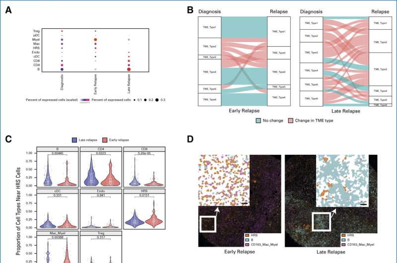
Distinct spatially resolved TME features according to relapse status. (A) Proportion for the indicated immune cell population by IMC-based cluster assignment. (B) The alluvial plot shows the TME types and their dynamic change between diagnostic samples and relapse samples according to relapse status. Horizontal ribbons represent individual cases and can be followed from left to right. Blue color of the ribbons indicates that there is no TME type change between diagnostic and relapse samples, whereas red-colored samples indicate the change in TME type. (C) Violin plot indicating the spatial score for the indicated cell types near HRS cells according to relapse status. (D) IMC analysis from FFPE sections of CHL shows localization of immune cells according to relapse status. (Left) A representative case with early relapse CHL case shows numerous CD163+ macrophage/myeloid cells and rare B cells. By contrast, (right) a representative late relapse HL case shows few CD163+ macrophage/myeloid cells and abundant B cells. (E) Box plot indicating the spatial scores of macrophage/myeloid cell subtypes near HRS cells according to relapse status. (F) Dot plot showing correlation of spatial scores of major immune cell markers by IMC. Dot size and color summarize Pearson correlation values, with positive correlations represented in red and negative correlations represented in blue. Asterisks represent associated P values (*P < .05; **P < .01; ***P < .001). cDC, conventional dendritic cell; CHL, classic Hodgkin lymphoma; FFPE, formalin-fixed paraffin-embedded; HRS, Hodgkin and Reed Sternberg; IMC, imaging mass cytometry; pDC, plasmacytoid dendritic cell; TME, tumor microenvironment; Treg, T regulatory cell. Credit: Journal of Clinical Oncology (2023). DOI: 10.1200/JCO.23.01115
Cedars-Sinai Cancer investigators have discovered a new way to predict whether a cancer of the immune system will recur in patients treated with a bone marrow transplant. Their study, published in the Journal of Clinical Oncology, is the first to use a novel technique called spatial profiling to predict patient outcomes, and could lead to more precisely targeted treatment.
"Our method of predicting how Hodgkin lymphoma patients will respond to treatment was more accurate than the most advanced method in current use," said Akil Merchant, MD, co-director of the Lymphoma Program at Cedars-Sinai and co-senior author of the study. "Our paper is also one of the first to show that this leading-edge technique can be adapted for a clinical setting—a finding that can potentially be used across cancer types."
Hodgkin lymphoma is a cancer that affects the lymphatic system, a network of organs and tissues—including the bone marrow, lymph nodes and mucous membranes—that helps protect the body from infection.
To treat Hodgkin lymphoma, doctors commonly perform a stem cell transplant, using healthy blood stem cells from the patient's own body, to help the bone marrow recover and to create new immune cells that can fight the cancer cells.
"Our new test, developed at Cedars-Sinai, allows us to identify a group of patients who will likely remain disease-free after this stem cell transplant," Merchant said. "For these post-transplant patients, the goal is to end subsequent treatments, sparing them from additional therapy with potentially life-threatening side effects. Our findings could also help us design clinical trials to identify therapies to help patients not cured by their transplant."
In this study, done in collaboration with Christian Steidl, MD, Ph.D., of British Columbia Cancer, investigators analyzed biopsies from 169 patients with Hodgkin lymphoma. They compared the cells and tissues immediately surrounding tumors from patients cured of bone marrow transplants with those from patients whose cancer returned following the transplant.
By looking at the distance between the cancerous cells and other cell types, they were able to predict how the cancer would respond to a transplant using the patient's own stem cells.
"Our study made use of massive datasets combined with machine learning that enabled us to focus on two or three key data points that could be used in a clinical test," Merchant said. "The test could be widely used to predict which patients are at high risk for relapse after transplant and allow clinicians to tailor their therapy accordingly."
Alexander Xu, Ph.D., research scientist at Cedars-Sinai Cancer and co-first author of the study, emphasized the value of such a quantitative tool. "Diagnosis and prediction often involve judgment calls. Because our study is quantitative, it will be important in maintaining consistent results from patient to patient and clinic to clinic," Xu said.
The investigators are exploring the creation of such a test. They are also pursuing projects to identify similar predictive tests for other types of cancer.
"This work opens new avenues for the development of spatial biomarkers that can guide treatment of cancer patients in a precise, targeted way," said Dan Theodorescu, MD, Ph.D., director of Cedars-Sinai Cancer and the PHASE ONE Distinguished Chair. "Translation of this leading-edge research to a test that can be clinically deployed will help bring the promise of precision medicine to increasing numbers of patients—and will eventually benefit patients with many cancer types."
More information: Tomohiro Aoki et al, Spatially Resolved Tumor Microenvironment Predicts Treatment Outcomes in Relapsed/Refractory Hodgkin Lymphoma, Journal of Clinical Oncology (2023). DOI: 10.1200/JCO.23.01115
Journal information: Journal of Clinical Oncology
Provided by Cedars-Sinai Medical Center


Post comments