
Reviewed by Lily Ramsey, LLM
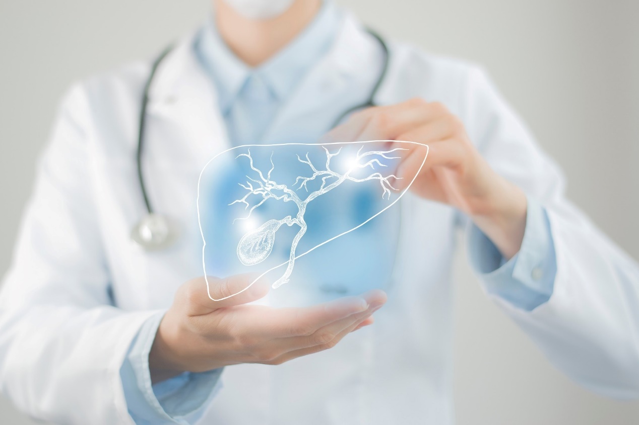
In a large, prospective study published in The Lancet, researchers from India developed and validated a deep-learning (DL) model to automate the detection of gallbladder cancer (GBC) via abdominal ultrasound (US) imaging.
The study found that with high sensitivity in performance comparable to radiologists, the DL-based method could detect GBC even in the presence of stones, in contracted gallbladders, with a lesion size less than 10 mm and neck lesions.
Further, the method's sensitivity for mural thickening type of GBC exceeded that of a radiologist.
Study: Deep-learning enabled ultrasound based detection of gallbladder cancer in northern India: a prospective diagnostic study. Image Credit: mi_viri/Shutterstock.com
Background
GBC is an aggressive biliary tract malignancy, mostly with a late-stage diagnosis, leading to a poor prognosis. It presents a diagnostic challenge as benign lesions present with similar imaging features.
Artificial intelligence (AI)-based approaches have recently revolutionized the radiological diagnosis of various cancers, potentially reducing human effort and improving sensitivity.
However, despite being a readily available, noninvasive imaging modality, US assisted by DL for diagnosing GBC has not been explored thoroughly.
Small sample sizes limited previous studies and did not evaluate the performance of DL models in diverse real-world scenarios. Therefore, this study aimed to train, validate, and test a DL algorithm based on imaging from a large dataset and compare its performance with that of radiologists to evaluate the potential of DL for GBC diagnosis.
About the study
In contrast to human experts' manual analysis, DL trains a computer to automatically recognize features and patterns in large datasets of sample images using convolutional neural networks (CNNs).
In this study, the researchers used a DL algorithm trained on a dataset of 565 prospective patients from northern India with gallbladder lesions acquired over a period of about two years. The mean age (±SD) was 50.8 ± 22.6 years, and 63.2% of the patients were females.
The exclusion criteria for the study were the presence of polyps ≤ five mm, acute cholecystitis, gallbladder abnormalities secondary to extra cholecystitis causes such as pancreatitis or hepatitis, and systematic illnesses like viral infections in patients.
Gallbladder US was performed on the Logiq S8 US scanner at 1–5 MHz after a minimum of six hours of fasting in supine and lateral decubitus positions. Patients with gallbladder polyps were assessed at higher frequencies, up to eight MHz.
Advanced DL techniques were used for GBC detection, including visual acuity-based learning and a publicly available, multiscale, second-order pooling-based classifier. The model's performance was assessed in a temporally independent test cohort and compared with the independent reviews of two experienced radiologists.
Specific criteria were used to diagnose GBC, benign lesions, or gallbladder wall thickening (GWT). Statistical analyses included determining the mean, 95% Clopper-Pearson confidence intervals, sensitivity, specificity, positive predictive value (PPV), negative predictive value (NPV), accuracy, and area under the receiver operating characteristic (ROC) curve (AUC) and comparison using the Mc Nemar test.
Results and discussion
The DL model showed 92.3% sensitivity, 74.4% specificity, 86.4% accuracy, and 0.887 AUC in the test cohort.
No significant difference was observed in the sensitivities, specificities, and AUCs of the model and those determined by the two radiologists, indicating the model's noninferiority in GBC detection.
As per the study, the model performed well in all the clinical subgroups, including distinct morphological subtypes, gallbladder lesions with stones, contracted gallbladders, lesions <10 mm, and distinct gallbladder sites.
Additionally, the DL model showed significantly higher sensitivity but reduced specificity than one of the radiologists when detecting GWT type of GBC (p=0.012).
These findings suggest that the DL model is a robust and promising option to improve GBC diagnosis's sensitivity, accuracy, and efficiency using US imaging in diverse clinical scenarios. This could prove particularly important in areas where access to specialized radiologists or advanced imaging techniques is limited.
However, the study is limited because only single-center data has been used, with a relatively smaller subgroup of patients with polyps. The impact of the method on early diagnosis and prognosis of GBC also remains to be studied.
Conclusion
This prospective study provides comprehensive evidence, on the largest-ever sample size, that the DL-assisted models can detect GBC in the presence of stones, contracted gallbladders, lesion size <10 mm, and neck lesions, which is challenging to diagnose conventionally.
The study findings demonstrate the potential utility of DL to improve the accuracy of GBC diagnosis and pave the way for further research in multicenter clinical settings. Furthermore, DL-assisted automatic GBC detection can help with early diagnosis and timely intervention, improving patient outcomes.
Journal reference:
Gupta P, et al., (2023). Deep-learning enabled US based detection of gallbladder cancer in northern India: a prospective diagnostic study. The Lancet (2023). doi:10.1016/j.lansea.2023.100279. https://linkinghub.elsevier.com/retrieve/pii/S2772368223001397


Post comments