
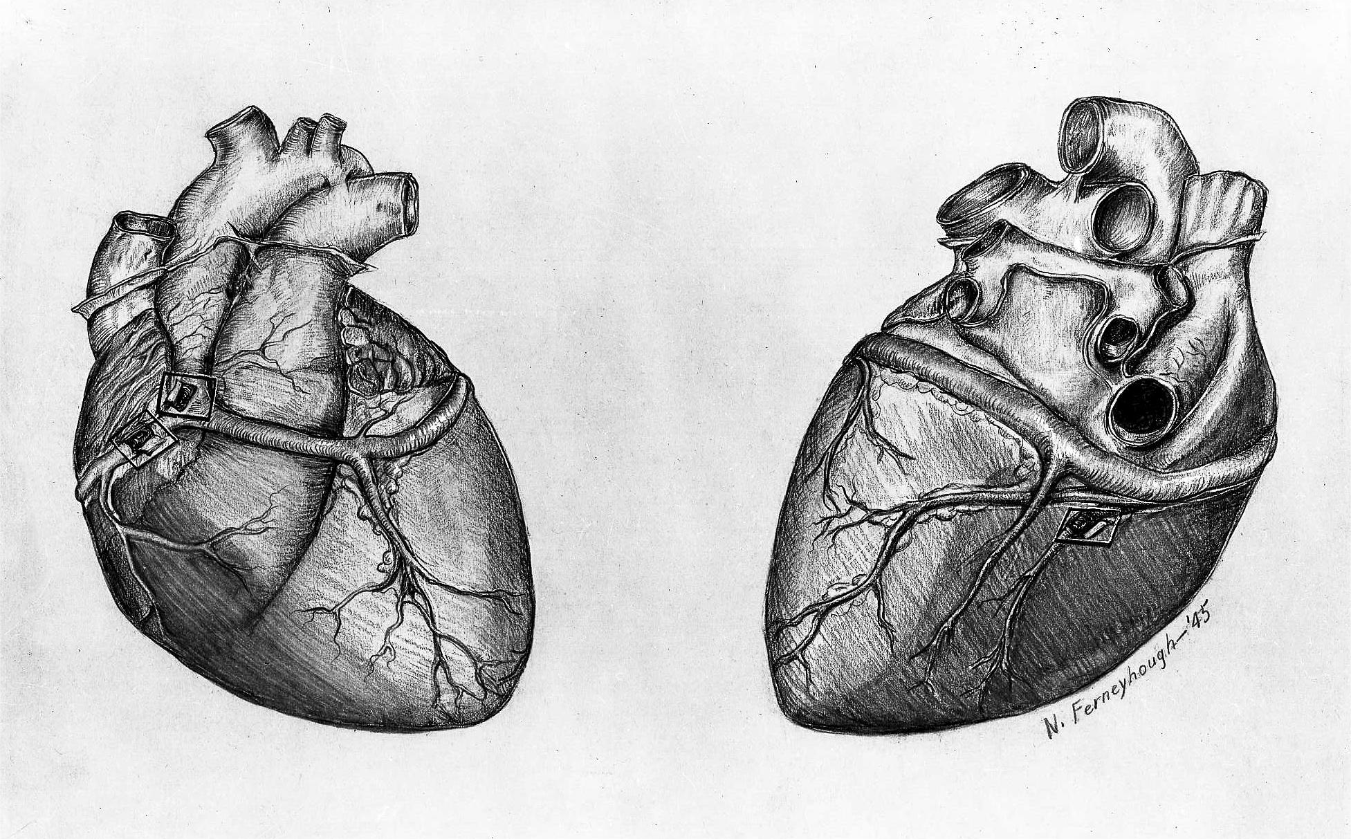
Credit:"Drawing heart (1945) N. Ferneyhough" is marked with CC0 1.0.
Heart Failure (HF) is a clinical condition characterized by impaired cardiac pump function, unable to meet the body's demand for blood and oxygen. Mitochondria play a crucial role in cardiac functions mainly through the oxidative phosphorylation process to produce ATP.
A recent study focuses on two specific enzymes—dual-specificity tyrosine-(Y)-phosphorylation regulated kinase 1B (DYRK1B) and signal transducer and activator of transcription 3 (STAT3), exploring how they influence mitochondrial bioenergetics to promote the occurrence of cardiac hypertrophy and heart failure. DYRK1B and STAT3 are two signaling molecules that play significant roles in cardiac diseases. They influence the survival, proliferation, metabolism, and function of cardiac cells by participating in various cellular signaling pathways. DYRK1B, a protein kinase, can phosphorylate various substrates, affecting the cell cycle, metabolism, and cell death, and it has been found to promote cardiac hypertrophy by regulating downstream signals such as NFAT and GSK-3β. Studies show that in heart failure models, increased expression of DYRK1B may exacerbate the pathology of heart disease by affecting the energy metabolism and survival rate of cardiac cells. Whereas STAT3, a transcription factor, is considered a protective signaling molecule in heart diseases, involved in various cellular functions including cell growth, differentiation, and immune responses, and its activation can reduce myocardial cell damage and improve cardiac function. Research data suggests that prolonged chronic cardiac stress leads to sustained activation of STAT3, which may help regulate the adaptive remodeling process of the heart, including promoting the survival of cardiac cells and reducing fibrosis.
The author generated genetically engineered mice specifically expressing DYRK1B and DYRK1B knockout mice, using aortic constriction surgery to establish a mouse model of cardiac hypertrophy. The study showed that compared to wild-type mice, the DYRK1B overexpressing mice exhibited significant declines in cardiac function, including reduced LVEF and thickening of the ventricular walls. DYRK1B knockout mice demonstrated better cardiac function than wild-type mice following TAC surgery, with less reduction in LVEF and less pronounced thickening of the ventricular walls, indicating a protective effect. Conversely, in DYRK1B knockout mice, the TAC surgery-induced increase in STAT3 phosphorylation was significantly inhibited, and the decrease in PGC-1α expression was partially reversed. Additionally, in the hearts of DYRK1B overexpressing mice, there was a noticeable increase in the size of cardiomyocytes and myocardial fibrosis, whereas these changes were significantly reduced in DYRK1B knockout mice.
These experimental results may validate the hypothesis that DYRK1B affects cardiac mitochondrial function and structure by regulating STAT3 and PGC-1α and its potential clinical meanings for further research on prevention and treatment of heart failure. It should be noted that this study primarily was on genetically modified mouse models, which may limit the generalizability of the results on human conditions. Therefore, the research findings need to be further validated in a broader range of animal models before considering clinical trials.
More information:
Zhuang L, Jia K, Chen C, Li Z, Zhao J, Hu J, Zhang H, Fan Q, Huang C, Xie H, Lu L, Shen W, Ning G, Wang J, Zhang R, Chen K, Yan X. DYRK1B-STAT3 Drives Cardiac Hypertrophy and Heart Failure by Impairing Mitochondrial Bioenergetics. Circulation. 2022 Mar 15;145(11):829-846.


Post comments