

Image by kjpargeter on Freepik
Heart failure is a critical condition that affects millions of people worldwide. Early identification of structural changes in the heart can significantly improve patient outcomes. However, the initial signs and symptoms of early-stage heart failure are often non-specific, leading to delays in diagnostic echocardiography. This delay can result in worse outcomes for patients. Therefore, finding an accurate, broadly applicable, and cost-effective method to detect left ventricular (LV) structural abnormalities is crucial. Recent advancements in deep learning have shown promise in addressing this challenge by utilizing chest X-rays (CXRs), a relatively inexpensive and frequently performed diagnostic tool.
Background and Aims
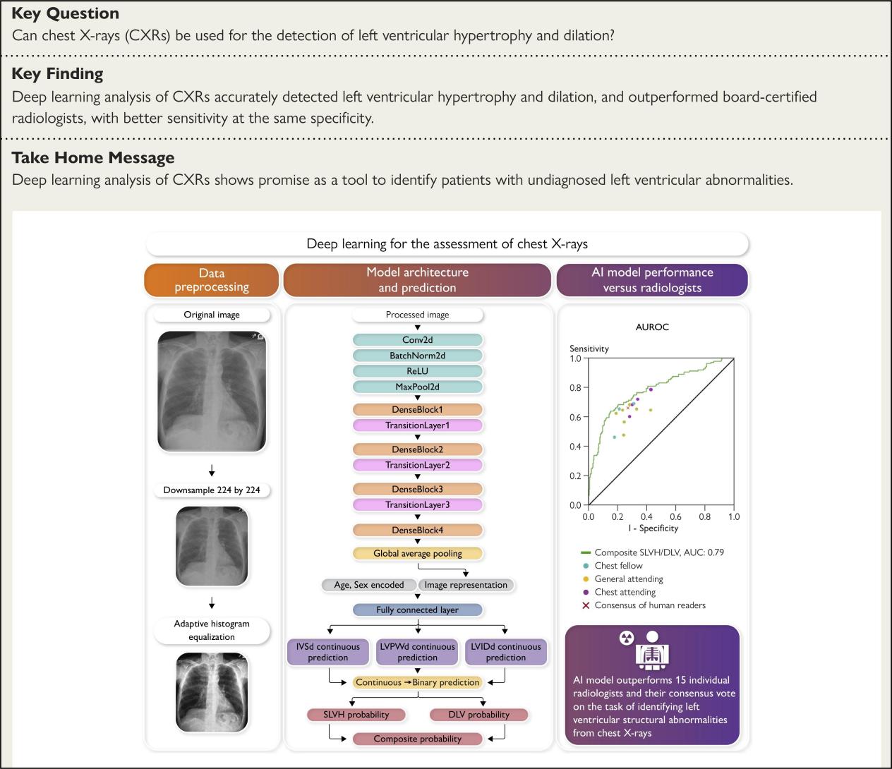
The primary aim of the study was to develop a deep learning model capable of identifying severe left ventricular hypertrophy (SLVH) and dilated left ventricle (DLV) using CXRs. These structural abnormalities are indicative of Stage B or worse heart failure. The study leveraged a large dataset of CXRs and echocardiograms to train and validate the model, with the goal of creating a scalable screening tool for early detection of heart failure.
Methods
The study identified 71,589 unique CXRs from 24,689 different patients, all completed within one year of echocardiograms. The echocardiographic measurements, including interventricular septal thickness at end-diastole (IVSd), LV internal diameter at end-diastole (LVIDd), and LV posterior wall distance at end-diastole (LVPWd), were used as gold-standard labels for training the model. The CXRs were pre-processed to ensure uniformity in size and contrast, and Gaussian noise was added to improve generalization performance.
The model architecture was based on DenseNet-121, a deep learning framework known for its effectiveness in learning representations from medical images. The final layer of the neural network combined the image representation with patient demographic information, such as age and sex, to make predictions.
Results
The model demonstrated strong performance in detecting SLVH and DLV, with an area under the receiver operating characteristic curve (AUROC) of 0.79 for SLVH, 0.80 for DLV, and 0.80 for a composite label indicating the presence of either condition. These results were consistent across an external dataset from Stanford University Medical Center, which included 8,003 CXRs from 4,657 patients.
In a head-to-head comparison with 15 board-certified radiologists, the model outperformed all individual radiologists in predicting the composite label. The model achieved a sensitivity of 71% compared to 66% for the consensus vote across all radiologists. This indicates that the deep learning model can detect structural abnormalities with higher accuracy than human experts.
Discussion
The study highlights the potential of deep learning models to serve as effective screening tools for early detection of heart failure. By leveraging CXRs, which are more commonly performed than echocardiograms, the model can identify patients with structural heart abnormalities who may benefit from further diagnostic evaluation. This approach aligns with recent guidelines from the European Society of Cardiology (ESC) and the American College of Cardiology (ACC), which advocate for more studies on screening for heart failure in asymptomatic patients.
One of the key strengths of the study is its use of high-quality labels derived from echocardiographic measurements. This ensures that the model is trained on accurate and clinically relevant data. Additionally, the study's methodology for evaluating the model on an external dataset and a subpopulation of patients who had CXRs prior to their first echocardiogram demonstrates the model's robustness and generalizability.
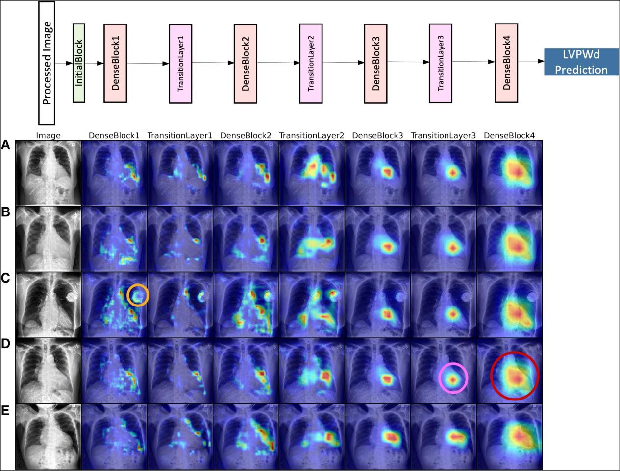
LayerCAM for chest X-rays with respect to left ventricular posterior wall distance at end-diastole continuous label. The figure shows saliency maps for five patients amongst the true positives for the composite label. For each patient, the feature map for each intermediate layer in the model architecture is displayed (the corresponding model layer is indicated above each column). The heatmaps show which areas of the image the model are attending to for making predictions. In analysing all heatmaps of the test set, broad patterns emerged represented by these examples: (orange circle, Patient C) in earlier layers the model can show sensitivity to implanted devices as highlighted in the orange outline for Patient C; (pink circle, Patient D) in intermediate layers, heatmaps are more concentrated within the borders of the cardiac silhouette and often highlighted areas corresponding to the left ventricle as shown in the pink outline; (red circle, Patient D) in the final layer, the model shows sensitivity to a broad area in the centre of the chest X-rays corresponding to the cardiac silhouette as shown in the red outline. LVPWd, left ventricular posterior wall distance at end-diastole.
Challenges and Future Directions
Despite the promising results, the study acknowledges several challenges. Differences in data distribution between institutions, variations in patient populations, and distinct practices related to reading echocardiograms can impact model performance. The study found that the model could accurately predict the source institution of the CXRs, indicating detectable differences in data distributions. Addressing these challenges will be crucial for ensuring the model's applicability across diverse clinical settings.
Future research should focus on further validating the model in larger and more diverse populations. Additionally, integrating the model into clinical workflows and assessing its impact on patient outcomes will be important steps toward translating this technology into practice. Explainability of deep learning models is also critical for building trust among clinicians. The study's use of saliency mapping to identify the parts of the CXRs most sensitive to the model's predictions is a step in this direction.
Conclusion
The development of a deep learning model to detect LV structural abnormalities from CXRs represents a significant advancement in the early diagnosis of heart failure. By providing a cost-effective and scalable screening tool, this approach has the potential to improve patient outcomes by enabling earlier intervention. As the field of artificial intelligence continues to evolve, such innovations will play an increasingly important role in transforming healthcare and enhancing the quality of care for patients with heart disease.
Read Paper ▏European Heart Journal


Post comments