
byCedars-Sinai Medical Center
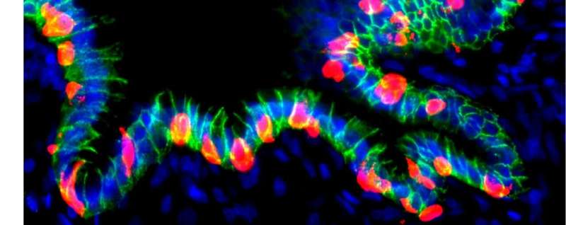
Credit:Diabetologia(2025). DOI: 10.1007/s00125-025-06558-5
A Cedars-Sinai study helps explain why half of diabetes patients experience deterioration of the cornea, the transparent dome-shaped outer layer of the eye that provides protection and focuses incoming light. The findings,publishedinDiabetologia, point to a potential target for therapies to protect vision in these patients.
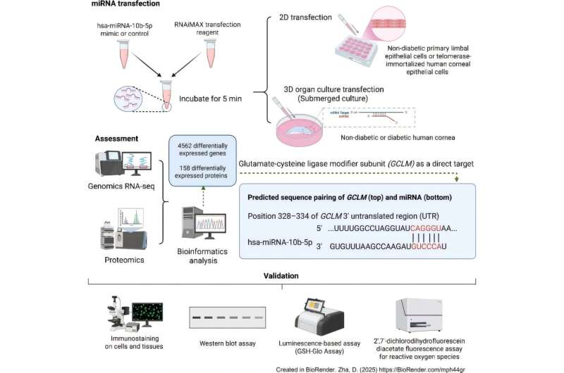
Investigators performed experiments comparing diabetic versus nondiabetic human corneal cells to explain the molecular changes that diabetespatientsexperience. Diabetic corneal disease can lead to delayed wound healing and loss of vision.
"Our findings highlight the major role of a molecule called microRNA-10b in the oxidative stress characteristic of diabetic corneal disease and the damage it causes to cells," said Mehrnoosh Ghiam, Ph.D., associate professor of Biomedical Sciences, research scientist in the Board of Governors Regenerative Medicine Institute and senior author of the study.
"Inhibiting this molecule restored the cornea's defenses againstoxidative stressandcell damageand allowed the outermost layer of cells to be maintained and renewed."
More information: Daxian Zha et al, Oxidative stress-regulatory role of miR-10b-5p in the diabetic human cornea revealed through integrated multi-omics analysis, Diabetologia (2025). DOI: 10.1007/s00125-025-06558-5 Journal information: Diabetologia
Provided by Cedars-Sinai Medical Center


Post comments