
by eLife
Credit: Pixabay/CC0 Public Domain
Scientists have found a potential new drug target for untreatable fatty liver disease in humans, according to a study published today in eLife.
Their findings, from mice and human liver biopsies, suggest that targeting a receptor found predominantly in the liver could help protect against the build-up of fat, halting the development or progression of non-alcoholic fatty liver disease (NAFLD).
NAFLD is a symptomless condition where fat builds up in the liver. It affects around 25% of the worldwide population and, if left untreated, accounts for approximately 85% of all chronic non-communicable diseases, such as type 2 diabetes, heart disease and chronic kidney disease. It can also lead to a condition called non-alcoholic steatohepatitis (NASH), the buildup of scar tissue in the liver (cirrhosis), and potentially liver cancer. It is caused by lipid metabolism disorders, over- or mal-nutrition, inflammation, viral infection or liver injury.
"Increasing physical activity and reducing calories can help to manage NAFLD but such lifestyle changes are only effective in its early stages," explains lead author Mengyao Wu, Guangzhou University, Guang Dong, China. "No medication is available to reverse the excessive fat storage in the liver once NASH has developed. Therefore, we urgently need to unravel the mechanisms of NAFLD to find targets for the development of diagnostic tests and cost-effective therapies."
Wong and colleagues focused their attention on a molecule found at high levels in the liver—the adhesion G-protein coupled receptor (Adgrf1), which was first identified in 2002. As a member of the G-protein coupled receptor (GPCR) family, it belongs to the largest and most diverse family of receptors on the cell surface, many of which are key targets for approved drugs. Although a few other GCPRs have been shown to play a role in NAFLD, they are not exclusively found in liver cells, and have the potential to cause side effects on other major organs. By contrast, Adgrf1 is found predominantly in the liver, but its function in this organ was yet to be determined.
In this study, Wong and colleagues first looked at the effect of a high-fat diet on the levels of Adgrf1 in the liver of mice. They found that Adgrf1 levels dramatically decreased in the liver cells of mice after a high-fat diet, which can be a novel marker for NAFLD.
Next, they looked at the effect of changing Adgrf1 levels on metabolism within liver cells. Overexpression of Adgrf1 had no significant effect on body weight, fasting glucose, fasting insulin or insulin resistance in lean subjects. But when Adgrf1 levels were not reduced after a high-fat diet, the mice had the features of diabetes—exhibiting higher fasting glucose and insulin levels, as well as insulin resistance.
To be certain that these changes were due to Adgrf1, the team used a gene therapy approach to block Adgrf1 in obese mice. In obese mice with fatty liver disease, suppressing Adgrf1 improved fasting glucose and insulin sensitivity. Moreover, blocking Adgrf1 also reduced lipid accumulation within liver cells, although circulating levels of fat in the blood remained high. Adgrf1-suppressing treatment also lowered levels of liver enzymes that are known markers of liver damage.
To work out how Adgrf1 has this effect, the team performed RNA sequencing on liver samples from mice to look at changes in gene expression. This revealed changes in an enzyme called stearoyl coA desaturase 1 (Scd1), a key fat-promoting enzyme known to play a role in NAFLD, circulatory diseases and diabetes. Their results suggested that Adgrf1 also plays a role in the synthesis of fat.
Next, they checked the relevance of their findings from mice in humans, using a public set of gene expression data for liver biopsy samples from patients at different levels of disease progression—from healthy liver through to NAFLD. Healthy (without NAFLD) obese people had lower Adgrf1 expression than healthy people who were not overweight, but surprisingly, people with NAFLD and obesity had similar Adgrf1 expression to lean people without NAFLD. Further analysis suggested that this surprising result was caused by inflammation in the livers of people with NAFLD, which increases levels of Adgrf1. They also found that Adgrf1 levels were higher in biopsies from patients with severe fatty liver disease (steatosis) than those with less severe NAFLD.
Taken together, these results suggest that reducing Adgrf1 expression could potentially serve as a protective mechanism to stop the overaccumulation of fat in the liver of people with obesity.
"We have found evidence for a novel role of Adgrf1 in regulating fat metabolism in the liver," concludes senior author Chi-Ming Wong, Associate Professor at The Hong Kong Polytechnic University, Hong Kong SAR, China. "The findings of our study pave the way for further research into the safety and efficacy of targeting Adgrf1 for the treatment of people with fatty liver disease. If confirmed, this could provide a new therapeutic approach for patients."
More information: Mengyao Wu et al, Amelioration of non-alcoholic fatty liver disease by targeting adhesion G protein-coupled receptor F1 (Adgrf1), eLife (2023). DOI: 10.7554/eLife.85131
Journal information: eLife
Provided by eLife
Estrogen-negative cancers respond to anti-estrogenic therapies, study shows
by Hokkaido University
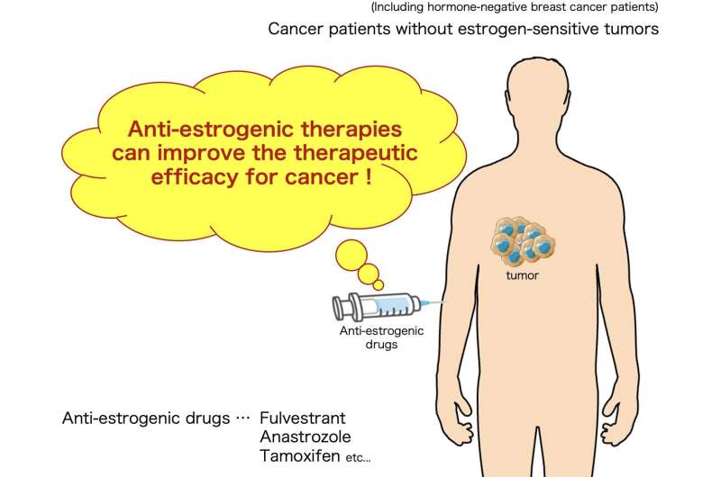
Anti-estrogenic therapies can suppress the growth of cancer that does not express estrogen receptors; when combined with immune checkpoint inhibitor therapies, they halt tumor progression in mice models.
Estrogen, a group of female hormones, is known to be involved in cancer progression, especially breast cancer. About 75% of breast cancers are estrogen-sensitive: they express the hormone receptor estrogen receptor α (ERα), and estrogen promotes tumor growth. Surprisingly, estrogen has been observed to promote tumor growth in ERα-negative cancers, such as triple-negative breast cancer (TNBC), for reasons that are not fully understood.
A team of researchers from the Institute for Genetic Medicine (IGM) at Hokkaido University has uncovered how estrogen affects the tumor microenvironment and promotes tumor growth in ERα-negative cancers. Their findings were published in the British Journal of Cancer.
"Generally speaking, estrogen directly affects cancer cells to promote cell survival and proliferation, and this has been considered to hold true only for estrogen-sensitive cancers," explains Nabeel Kajihara, lead author of the paper. "Estrogen is also documented to play other roles in the tumor microenvironment, effectively suppressing the immune response and protecting tumors."
The researchers analyzed patient data from The Cancer Genome Atlas (TCGA) and conducted experiments in cell cultures and mice models to understand what was happening.
From the TCGA data, they observed that, in TNBC, estrogen suppresses the induction of cytotoxic T cells, which typically recognize and destroy cancer cells. They confirmed this observation in mice TNBC and colon cancer models, which do not have estrogen sensitivity; they then went one step further and studied the effects of anti-estrogenic therapy on these cancers in mice models.
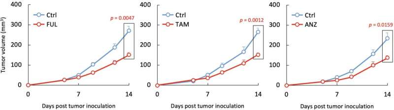
Therapy with fulvestrant, the most effective estrogen signal blocker currently approved for clinical use, suppressed the growth of tumor cells. Tumor growth was also suppressed by two other approved anti-estrogenic drugs, tamoxifen and anastrozole, confirming that estrogen signal-blocking was responsible.
Estrogen signal blockers modulate immune response to increase production of cytotoxic T cells, specifically by preventing the activity of estrogen. The team also showed that therapy combining estrogen signal blockers with a class of drugs called immune checkpoint inhibitors (ICIs) drastically suppresses tumor progression in mice models. Most notably, the combination of fulvestrant (anti-estrogenic) and anti-CTLA-4 (ICI) resulted in the complete suppression of TNBC tumor progression.
"We have demonstrated that anti-estrogenic therapies can potentially maximize therapeutic efficacy of cancer immunotherapy regardless of tumor cells' ERα expression," concludes Professor Seino. "However, this is only the first step; future clinical research is required to develop cancer therapies that utilize this approach."
More information: Nabeel Kajihara et al, Blocking of oestrogen signals improves anti-tumour effect regardless of oestrogen receptor alpha expression in cancer cells, British Journal of Cancer (2023). DOI: 10.1038/s41416-023-02381-0
Journal information: British Journal of Cancer
Provided by Hokkaido University


Post comments