
by American Roentgen Ray Society
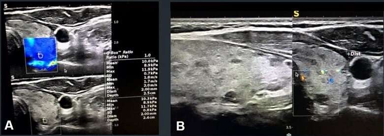
A. Elastography shows mostly blue pattern; B. Doppler sonography shows no flow pattern. Credit: ARRS
A scientific online poster presented findings that assessing the vascularity and elastography in suspect TIRADS categories can efficiently diagnose malignancy of thyroid nodules. This study was presented at the 2023 ARRS Annual Meeting on the island of Oahu.
Acknowledging that sonographic TIRADS scoring remains the first method of imaging assessment for diagnosing malignant thyroid nodules, "we assessed the added value of shear-wave elastography (SWE) to classic TIRADS assessment," said Leila Aghaghazvini, MD, from the department of radiology at Shariati Hospital and Iran's University of Medical Sciences in Tehran.
In this ARRS Annual Meeting Cum Laude award-winning online poster, 200 total thyroid nodules were assessed by conventional sonography to ascertain TIRADS score and pertinent descriptors. Using a 7.5 MHz probe to asses vascularity pattern and resistive index (RI), elastography evaluations were performed quantitatively via color mapping and calculating mean and maximum velocities in shear-wave modality. Final diagnoses of all thyroid nodules were confirmed by histopathology assessment or follow-up imaging.
According to Aghaghazvini and colleagues, patient mean age was 53 years; 105 were men [52.5%]. And of 27 [13.5%] total nodules with malignancy in their study, 14 [7%] were papillary thyroid carcinoma, 11 [5.5%] were follicular thyroid carcinoma, and 2 [1%] were medullary thyroid cancer. AUC of the ROC curve for TIRADS assessment was 0.76. Results found 37 [18.5%] TIRADS 4 nodules, wherein 15 [40.5%] were malignant, and 57 [28.5%] TIRADS 5 nodules, 12 [21.1%] of which were malignant.
In only TIRADS 4 nodules, Doppler grade AUC, RI, color map elastography grade, SWE maximum velocity, and mean SWE velocity for diagnosing malignancy were 0.8, 0.93, 0.89, 0.86, and 0.82, respectively. Meanwhile, profiles in TIRADS 5-only nodules were 0.8, 0.96, 0.85, 0.96, and 0.97, respectively. Additionally, in TIRADS 4, an RI cutoff point of 0.6 yielded diagnostic efficacy indices of 0.93, 1, 0.97, 1, and 0.96 for sensitivity, specificity, efficiency, PPV and NPV, respectively. In TIRADS 5 nodules, a cutoff point of 4.33 for SWE mean velocity yielded diagnostic efficacy indices of 1, 0.82, 0.86, 0.6, and 1 for sensitivity, specificity, efficiency, PPV and NPV, respectively.
Provided by American Roentgen Ray Society


Post comments