
by Sanford-Burnham Prebys
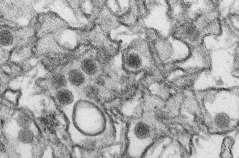
Transmission electron micrograph (TEM) of Zika virus. Credit: Cynthia Goldsmith/Centers for Disease Control and Prevention
Viruses have limited genetic material—and few proteins—so all the pieces must work extra hard. Zika is a great example; the virus only produces 10 proteins. Now, in a study published in the journal PLOS Pathogens, researchers at Sanford Burnham Prebys have shown how the virus does so much with so little and may have identified a therapeutic vulnerability.
In the study, the research team showed that Zika's enzyme—NS2B-NS3—is a multipurpose tool with two essential functions: breaking up proteins (a protease) and dividing its own double-stranded RNA into single strands (a helicase).
"We found that Zika's enzyme complex changes function based on how it's shaped," says Alexey Terskikh, Ph.D., associate professor at Sanford Burnham Prebys and senior author of the paper. "When in the closed conformation, it acts as a classic protease. But then it cycles between open and super-open conformations, which allows it to grab and then release a single strand of RNA—and these functions are essential for viral replication."
Zika is an RNA virus that's part of a family of deadly pathogens called flaviviruses, which include West Nile, dengue fever, yellow fever, Japanese encephalitis and others. The virus is transmitted by mosquitoes and infects uterine and placental cells (among other cell types), making it particularly dangerous for pregnant women. Once inside host cells, the virus re-engineers them to produce more Zika.
Understanding Zika on the molecular level could have an enormous payoff: a therapeutic target. It would be difficult to create safe drugs that target the domains of the enzyme needed for protease or helicase functions, as human cells have many similar molecules. However, a drug that blocks Zika's conformational changes could be effective. If the complex can't shape-shift, it can't perform its critical functions, and no new Zika particles would be produced.
An efficient machine
Researchers have long known that Zika's essential enzyme was composed of two units: NS2B-NS3pro and NS3hel. NS2B-NS3pro carries out protease functions, cutting long polypeptides into Zika proteins. However, NS2B-NS3pro's abilities to bind single-stranded RNA and help separate the double-stranded RNA during viral replication were only recently discovered.
In this study, the researchers leaned on recent crystal structures and used protein biochemistry, fluorescence polarization and computer modeling to dissect NS2B-NS3pro's life cycle. NS3pro is connected to NS3hel (the helicase) by a short amino acid linker and becomes active when the complex is in its closed conformation, like a closed accordion. The RNA binding happens when the complex is open, whereas the complex must transition through the super-open conformation to release RNA.
These conformational changes are driven by the dynamics of NS3hel activity, which extends the linker and eventually "yanks" the NS3pro to release RNA. NS3pro is anchored to the inside of the host cell's endoplasmic reticulum (ER)—a key organelle that helps shepherd cellular proteins to their appropriate destinations—via NS2B and, while in the closed conformation, cuts up the Zika polypeptide, helping generate all viral proteins.
On the other side of the linker, NS3hel separates Zika's double-stranded RNA and conveniently hands a strand over to NS3pro, which has positively charged "forks" to grab on to the negatively charged RNA.
More information: Sergey A. Shiryaev et al, Dual function of Zika virus NS2B-NS3 protease, PLOS Pathogens (2023). DOI: 10.1371/journal.ppat.1011795
Journal information: PLoS Pathogens
Provided by Sanford-Burnham Prebys


Post comments