
Credit:https://doi.org/10.1016/j.bone.2024.117110.
IL-1 is a highly active cytokine and a potent agonist capable of stimulating a variety of biological responses.IL-1 is secreted by the vast majority of nucleated cells, but its primary source is monocyte macrophages, followed by epithelial cells, chondrocytes, and synovial cells. IL-1 is categorized into two types, IL-1α and IL-1β. These two subtypes have different secretion mechanisms, in which IL-1α is mainly involved in IL-1 autocrine secretion within the cell in the form of precursors, and IL-1β is transported to bind to the cell membrane and participate in paracrine secretion. IL-1β precursors, on the other hand, mainly undergo cleavage by IL-1 converting enzyme and are transported to the extracellular compartment so as to fulfill their functions [1].
IL-1β is highly expressed in synovial fluid and has a significant correlation with cartilage damage. IL-1 is highly expressed in OA chondrocytes and matrix. It has been shown that the cell membrane proteins of OA chondrocytes contain a large number of IL-1 receptors, and their expression is twice that of normal chondrocytes. The main mechanisms of IL-1 in OA chondropathy are to promote chondrocyte apoptosis, inhibit cartilage matrix formation, induce up-regulation of matrix metalloproteinases (MMPs), and participate in the expression of inflammatory mediators. In addition, synoviocytes are also highly sensitive to IL-1, which can promote synoviocytes to express excessive PGE2, thus aggravating synovial inflammation [2].
Chondrocytes are an important component of articular cartilage. Generally, their growth and apoptosis are in dynamic balance. However, in the presence of IL-1, chondrocytes can produce large amounts of nitric oxide (NO), and excessive NO can cause apoptosis of chondrocytes and lead to degradation of the cartilage matrix. At the same time, NO can also weaken the matrix's ability to resist trauma. In addition, it has been shown that Fas, the membrane receptor for apoptosis, induces cell death when it binds to its ligand FasL. In the joint fluid of patients with OA, overexpression of FasL also induces chondrocyte apoptosis. IL-1 stimulation of chondrocytes is able to activate the Fas pathway, which ultimately leads to chondrocyte apoptosis [3].
Articular cartilage is composed primarily of extracellular matrix, and when matrix degradation exceeds synthesis, leading to cartilage destruction and ultimately aggravation. MMPs are widely found in connective tissues and consist of more than 20 members that capable of remodeling tissues. In addition, MMPs degrade type II collagen and proteoglycans, destroying articular cartilage and exacerbating the progression of OA. Endogenous tissue inhibitors of metalloproteinases (TIMPs), the main inhibitors of the MMPs family, are also found in the human body. On the one hand, IL-1 accelerates the synthesis of MMPs; on the other hand, IL-1 also inhibits the production of TIMPs, leading to an elevated ratio of the two, accelerating matrix degradation and ultimately leading to OA [4].
OA is an aseptic, chronic synovitis that is often accompanied by inflammatory signs and symptoms such as joint pain and swelling. IL-1 also plays an important role in synovial inflammation, and its expression is significantly up-regulated in OA synovial tissues and in borderline synovial tissues after arthroplasty [5]. In the early stage of OA, synovitis is closely related to intrinsic immunity, and IL-1β can prompt synovial cells to secrete large amounts of pro-inflammatory factors, such as PGE2 and ICAM-1, leading to an inflammatory response in the synovium. In addition, the newly produced PGE2 is able to promote the breakdown of cartilage, further aggravating the development of OA inflammation [6].
Reference:
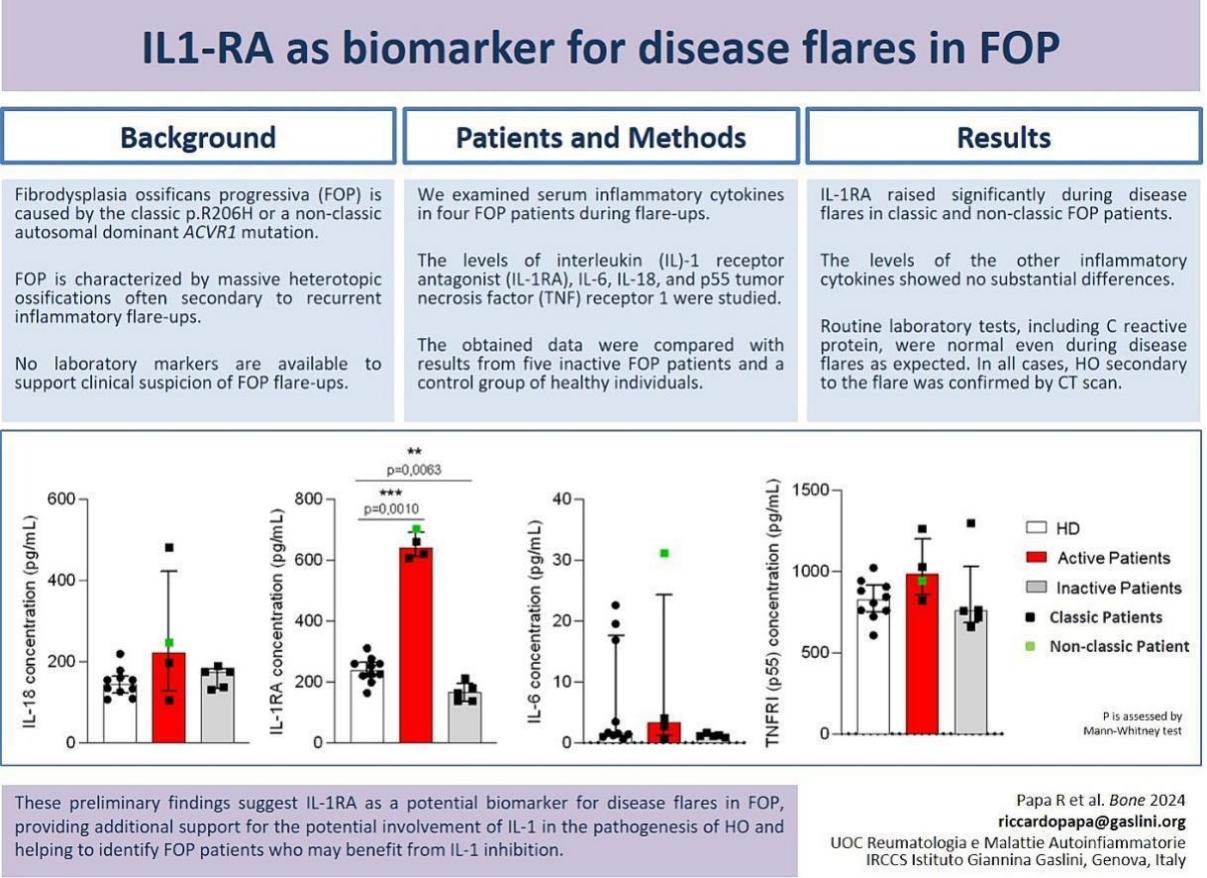
Papa R, Bertoni A, Matucci-Cerinic C, Drago E, Liberatore F, Corcione A, Gattorno M. Interleukin 1 receptor antagonist as biomarker for disease flares in fibrodysplasia ossificans progressiva. Bone. 2024 Jul;184:117110.
Adachi M, Okamoto S, Chujyo S, Arakawa T, Yokoyama M, Yamada K, Hayashi A, Akita K, Takeno M, Itoh S, Takii T, Waguri-Nagaya Y, Otsuka T, Hayakawa K, Miyazawa K, Onozaki K. Cigarette smoke condensate extracts induce IL-1-beta production from rheumatoid arthritis patient-derived synoviocytes, but not osteoarthritis patient-derived synoviocytes, through aryl hydrocarbon receptor-dependent NF-kappa-B activation and novel NF-kappa-B sites. J Interferon Cytokine Res. 2013 Jun;33(6):297-307.
Wang J, Chen L, Jin S, Lin J, Zheng H, Zhang H, Fan H, He F, Ma S, Li Q. Altered expression of microRNA-98 in IL-1β-induced cartilage degradation and its role in chondrocyte apoptosis. Mol Med Rep. 2017 Sep;16(3):3208-3216.
Cai S, Zheng J, Song H, Wu H, Cai W. Relationship between serum TGF- β 1, MMP-9 and IL-1β and pathological features and prognosis in breast cancer. Front Genet. 2023 Jan 12;13:1095338.
Ma T, Wu CB, Shen QX, Wang Q, Zhou Q. TRIM52 knockdown inhibits proliferation, inflammatory responses and oxidative stress in IL-1β-induced synovial fibroblasts to alleviate temporomandibular joint osteoarthritis. J Cell Mol Med. 2024 Apr;28(8):e18244.
Mao M, Li Y, Wang L, Chen J, Ming X, Sun Y, Lu Y, Ning J. Aitongxiao improves pain symptoms of rats with cancer pain by reducing IL-1, TNF-α, and PGE2. Int J Clin Exp Pathol. 2021 Jan 1;14(1):133-139.


Post comments