
by Brigitte Blöchlinger, University of Zurich
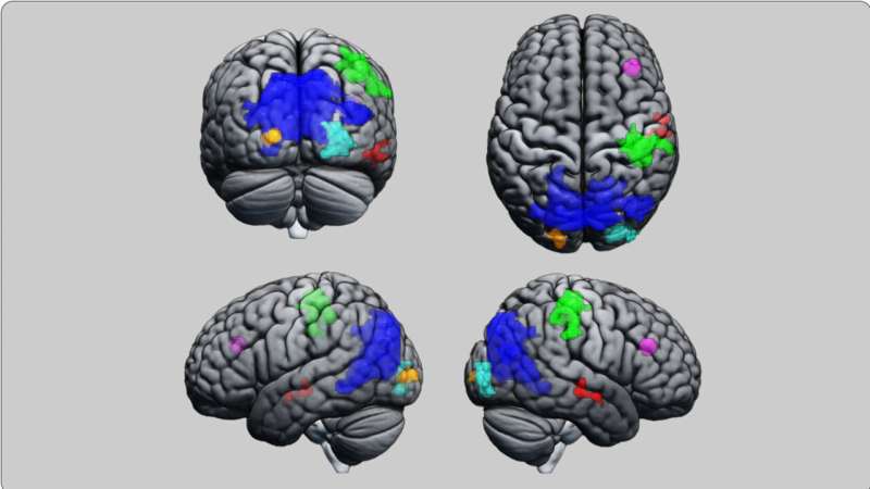
fMRI images show in color those regions of subjects’ brains where changes to functional connectivity were found at hypnosis state HS 2 (clockwise: brain from the rear, top, left and right). Credit: fMRI study, Mike Brügger, UZH
Three studies at the University of Zurich demonstrate that hypnosis alters activity in the large-scale functional networks of the brain. It also affects the neurochemical milieu of specific brain areas.
Hypnosis has so far been something of a black box from the scientific perspective. Up to now, we have not had the data to prove whether hypnosis really is an extraordinary state of human consciousness, or simply in the subject's imagination. Yet it remains a topic of fascination for many.
A well-known women's magazine recently dedicated an entire dossier to hypnosis. And now and again we'll hear of a remarkable hypnosis success story. For example, in 2018 at the Hirslanden Klinik St. Anna in Lucerne, a 45-year-old man had a metal plate removed from his lower arm under hypnosis only, without any anesthetic or pain relief. Much to the amazement of the surgical team, the man did not experience any significant pain either during or after the operation, as the Swiss public broadcaster SRF Puls health magazine program reported on 17 September of that year.
Alleviating pain and anxiety
Experience with hypnosis in a day-to-day hospital setting also seems to be very positive. The Geneva University Hospitals (HUG) offer clinical hypnosis as a complement to conventional treatment pathways. HUG also trains its staff accordingly, as it reports on its website. Furthermore, it occasionally successfully uses hypnosis with children who are afraid of an upcoming procedure, to ease their anxiety ahead of an examination.
Hypnosis seems to alleviate pain and fear. It is therefore sometimes used in ante-natal preparation, for burns or at the dentist.
Altered consciousness
Up to now, the exact impact of hypnosis on the human neurobiological system has remained unknown. Can changes to the brain be observed under hypnosis and, if so, what are they? These were the questions that neuroscientists Philipp Stämpfli, Nuno Prates de Matos and Mike Brügger set out to answer with other researchers in three studies. Two of these were conducted at the Center for Magnetic Resonance Imaging at the University Hospital of Psychiatry Zurich (PUK), which Stämpfli heads. The other study was held in the Department of Psychology of UZH.
Researching and describing different states of human consciousness is still one of the greatest challenges in neurobiology. It can be assumed that changes in states of consciousness will also present in the functional networks in the brain. Researchers therefore hypothesized that any effects of hypnosis would show up in these networks, and that it must be possible to map and measure them using imaging technology.
Three standardized studies
The neuroscientists conducted the three identically designed studies using different imaging methods in each case. This allowed them to analyze the effect of hypnosis on the human brain via a multimodal approach. The researchers believe that it was the first scientific project on hypnosis in the world to be so standardized and multimodal. It also studied two different depths of hypnosis for the first time.
The fMRI study was published in Frontiers in Human Neuroscience, the EEG study was published in Cortex, and the magnetic resonance spectroscopy study was published in Scientific Reports.
On each occasion, researchers selected just over 50 people as subjects. All were healthy, hypnosis experienced, and familiar with both hypnotic states. Once in the MR scanner, they were first brought into a mildly hypnotized state (HS1, somnambulism) using the same, standardized spoken text. They were then put into a very deep state of hypnosis (HS2, Esdaile).
Since the subjects were all experienced in hypnosis, they were able to give the study leads an agreed signal from the scanner once they had reached the relevant depth. They remained in this state for around 20 minutes, which is as long as it took to examine their brain with one of the three different imaging methods.
The researchers are aware that the sample for the three studies was highly selective. For that reason, they are keen to emphasize that their findings cannot be generalized. Instead, says Mike Brügger, "we wanted to determine basic principles, in other words whether there are differences in the brain when people were hypnotized to two different depths."
The aim of the three hypnosis studies was thus to gain a fundamental understanding of what happens during hypnosis, and not to investigate hypnosis as a possible form of treatment.
Network connectivity also changes
The three studies show from differing methodological perspectives and verifiable data that scientists can distinguish between two depths of hypnosis.
The fMRI study revealed changes in activity among the regions of the cerebral cortex that are involved in processes relating to attention and bodily awareness. All of the subjects reported feeling a very deep state of relaxation that was associated in some cases with a loss of perception of space and time.
Theta brainwaves increased at both levels of hypnosis. Theta waves are a sign of sleepiness and profound relaxation, and present additionally with other deep relaxation processes such as meditation or psychedelic states. They are also known to occur at different phases of the sleep cycle, but are not dominant during sleep.
In fact, the subjects also reported that, although they were very relaxed, they had been a long way away from actually falling asleep. Instead, researchers found that they tended to be highly focused.
Breathing and heart rate also slowed in subjects under hypnosis—another indicator of a state of deep relaxation.
Insights and outlook
The three studies have gathered an enormous amount of scientific data, but interpreting it is difficult, as the researchers freely admit. Despite this, Stämpfli and Brügger say that the studies have already answered two questions.
The first is that hypnosis genuinely does change something in the brain. The hypnotic effect is neither invented nor feigned. The second is that there is more than one depth of hypnosis. "That has been a really contentious issue in professional circles up to now, and there was little scientific evidence for it," according to Stämpfli.
These latest findings are a step forward, but the researchers are clear that further studies are needed to gain a general understanding of how hypnosis works. The question of why hypnosis can have a therapeutic effect on conditions such as pain or anxiety must also be explored in more depth.
The three imaging methods
Researchers used three different measuring methods for the three hypnosis studies.
They used electroencephalography (EEG) to track changes in brainwave frequency bands and spatial patterns in the collective electrical activity of millions of nerve cells. They applied functional magnetic resonance imaging (fMRI) to identify changes in communication between areas of the brain and present them in image form.
Building on these fMRI findings, they were able to use magnetic resonance spectroscopy (MRS) to investigate key areas of the brain at a deeper, molecular level.
More information: Nuno M. P. de Matos et al, Investigating functional brain connectivity patterns associated with two hypnotic states, Frontiers in Human Neuroscience (2023). DOI: 10.3389/fnhum.2023.1286336
Maria Niedernhuber et al, An interhemispheric frontoparietal network supports hypnotic states, Cortex (2024). DOI: 10.1016/j.cortex.2024.05.008 EEG
Nuno Miguel Prates de Matos et al, Neurochemical dynamics during two hypnotic states evidenced by magnetic resonance spectroscopy, Scientific Reports (2024). DOI: 10.1038/s41598-024-80795-3
Journal information: Frontiers in Human Neuroscience , Scientific Reports , Cortex
Provided by University of Zurich


Post comments