
By Peter Pressman, MD
Medically reviewed by Nicholas R. Metrus, MD
A brain tumor can be diagnosed using imaging tests that view the structure of the brain, along with a biopsy, which can carefully assess a sample of a suspected brain tumor under a microscope. Generally, before these tests are ordered, a physical examination is done to determine whether there are neurological changes that suggest the presence of a brain tumor. In the end, diagnosis of a brain tumor can involve an MRI, CT scan, blood tests, lumbar puncture, and biopsy.
There are several types of brain tumors, and some are cancer, which grows quickly and can invade nearby tissue, while some are not. These diagnostic tests can help a healthcare provider tell whether or not a person has a brain tumor and, if present, what type of brain tumor it is.
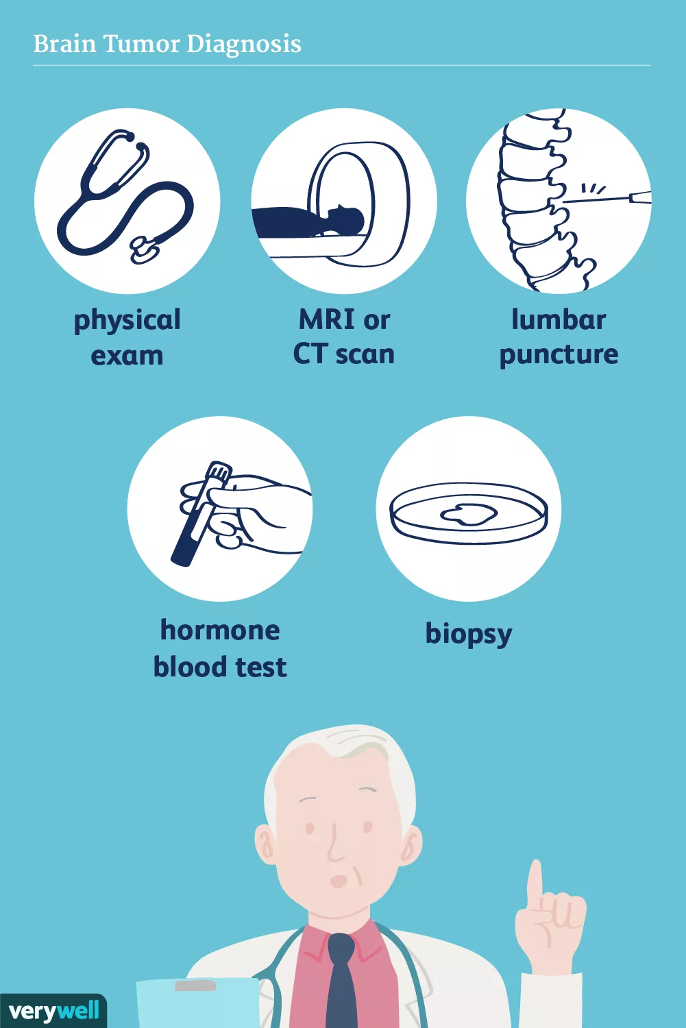
Illustration by Verywell
Self-Checks
A brain tumor is located inside the skull, so there are generally no changes that you are able to see on your own. However, there are a few signs of brain tumors that you should be aware of, especially because they can be subtle and slowly progressive.
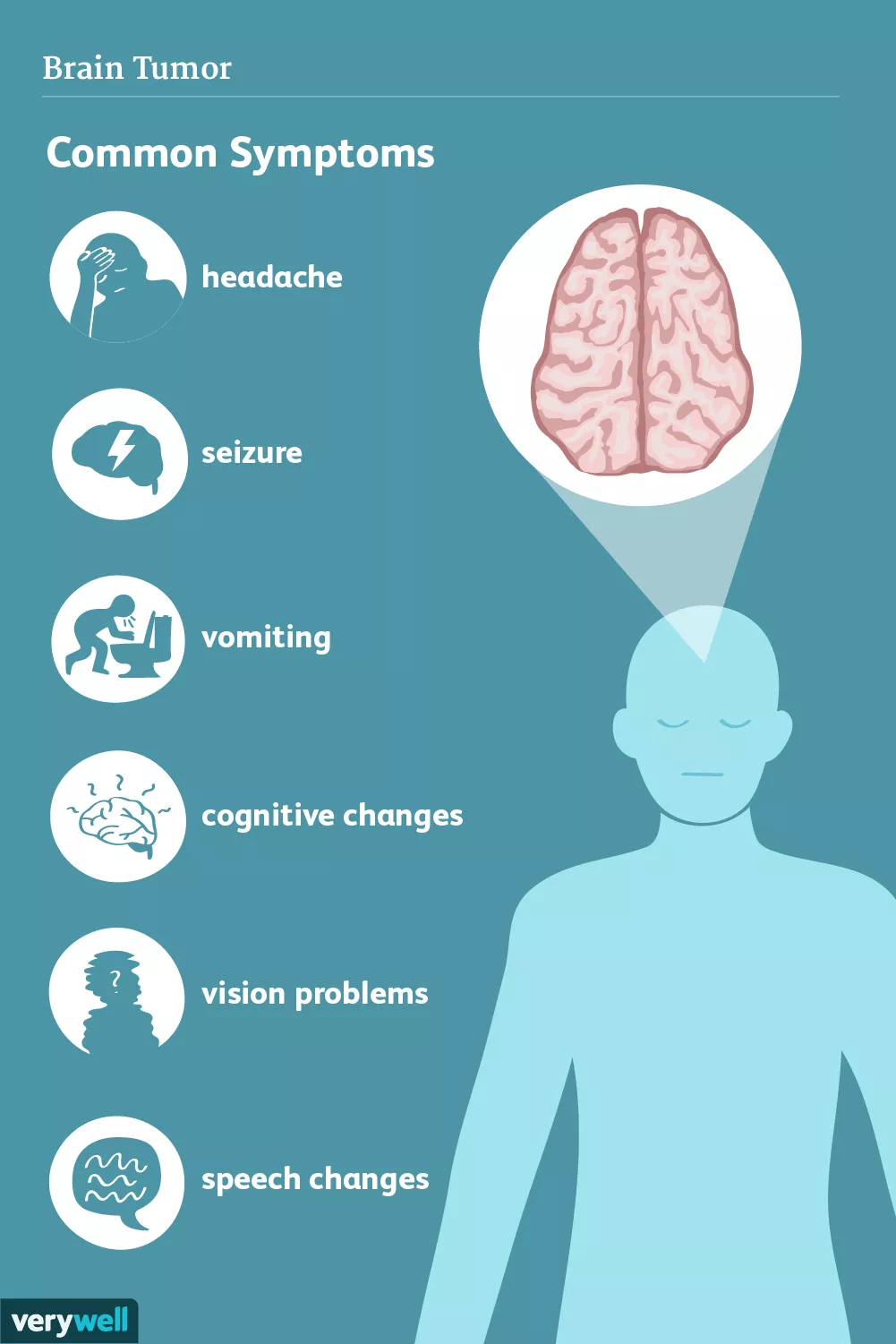
Be sure to take note of any of the following and bring them to the attention of your healthcare provider:1
Persistent headaches*
Vision changes
Coordination problems, such as an inability to stand up straight or difficulty using one of your hands
Unexplained vomiting
Weakness, numbness, tingling of the arms or legs
Difficulty speaking or understanding speech
Seizures
*While persistent headaches can be a sign of a brain tumor, they—in the absence of other symptoms—are rarely owed to one. Still, see your healthcare provider.
Physical Examination
Your healthcare provider's physical examination can help determine whether you have a possible brain tumor.
In general, neurological abnormalities that correspond to a section of the brain are associated with brain tumors, while those that correspond to an artery in the brain are caused by a stroke. These subtle differences can help your neurologist or neurosurgeon efficiently plan your diagnostic workup so that you can get the right diagnosis sooner.
Signs of a brain tumor on a physical examination can include weakness as well as a tremor, coordination problems on both sides of your body, or jerking movements of your eyes.
Most importantly, close examination of your eyes using an ophthalmoscope can reveal swelling, which is evidence of increased pressure in the brain caused by a brain tumor.
Imaging
Imaging can assess the size of a tumor and its location within the brain, as well as characteristics that help to differentiate one type of tumor from another.
For example, brain metastases tend to be located near small blood vessels, where tumor cells are more likely to cross the blood-brain barrier. Another type of brain tumor, glioblastoma multiforme, tends to be a large tumor that spreads across several different areas of the brain. A brain tumor called an oligodendroglioma may have bright spots on a brain CT scan due to calcium deposits within the brain.
The most common imaging tests for brain tumors are magnetic resonance imaging (MRI) and computed tomography (CAT scan, CT scan).2 These tests are usually done with injected contrast material, which is fluid that surrounds solid areas, such as brain tumors, to better define the edges.
Other tests often used for research purposes and sometimes surgical planning include magnetic resonance spectroscopy (MRS) and functional MRI (fMRI), which detect differences in metabolic activity that may occur with a brain tumor. A diffusion-weighted image uses MRI linked to software that calculates changes in the diffusion of water, which also may be altered when someone has a brain tumor.
Similarly, a positron emission test (PET) is similar to a CT scan and can detect microscopic changes in blood flow and oxygen consumption, which may occur with some brain tumors.
These types of imaging tests may not be available in every hospital, and the results are not considered as reliable or consistent in brain tumor diagnosis as contrast-enhanced brain CT or brain MRI, but they are valuable because they detect subtle changes that scientists use to learn more about brain disease.
Some other imaging tests can be used in surgical planning. For example, an angiogram uses CT, ultrasound or MRI to observe blood vessels, and can be used so that your surgeon can see if the tumor is near a blood vessel.
Some of the most common types/descriptions of tumors include:3
Primary brain tumor: A primary brain tumor is a tumor that started in the brain. It may grow, spread, or stay in one small spot, depending on which type of brain cell the primary brain tumor originally started from.
Metastatic brain tumor: A metastatic tumor in the brain is one that started somewhere outside the brain, such as the breast, lungs, or colon, and spread to the brain. Generally, a metastatic tumor is a highly aggressive tumor that might not improve with treatment.
Meningioma: This is a primary brain tumor that grows from the meninges, the protective tissue that surrounds the brain, and not actually from the brain itself. Meningioma is among the most common types of brain tumors. Depending on the grade of a meningioma, which is determined with a biopsy, it may have a good prognosis if it is completely removed surgically, or there may be a chance of recurrence or invasion of the brain.
Pituitary tumor: Another primary brain tumor that can be either completely treatable or very aggressive, a pituitary tumor is cancer of the pituitary gland, a structure in the brain that controls hormones. As with other brain tumors, a pituitary tumor can be visualized on brain imaging studies, and unlike other brain tumors, it can produce hormonal changes that can have a wide-ranging impact on the body.
Glioma: A primary brain tumor that arises from glial cells, which are supportive nerve cells in the brain, a glioma can begin on the optic nerve, the brainstem, or the cerebral cortex. Gliomas tend to be highly aggressive tumors, requiring intense treatment.
Labs and Tests
Blood tests can also help in assessing some types of brain tumors, and a lumbar puncture may help in diagnosing metastatic (aggressively spreading) tumors in the brain. A biopsy is a major procedure, and it is the most definitive test for brain tumor diagnosis.
Hormone Blood Tests
Some brain tumors, such as pituitary tumors, can produce hormones that are detected in the blood. If you have a pituitary tumor, you may have an abnormal concentration of hormones such as growth hormone or thyrotropin (a hormone that stimulates the thyroid gland) in your blood.4 These are not routine tests, so your healthcare provider would only order them if there is a high suspicion of a hormone-producing brain tumor.
Lumbar Puncture (LP)
For this test, commonly referred to as a spinal tap, a healthcare provider extracts fluid from your lower spine using a needle, which is then tested. It can help identify infections, inflammation, or cancer cells.
Cancer cells can appear in the cerebrospinal fluid (CSF) if you have carcinomatosis—a condition in which multiple areas of one organ are affected by metastatic cancer.5 Carcinomatosis in the brain can occur due to cancer that started somewhere else in the body or due to the spread of brain cancer within the brain.
However, LP is not usually a reliable test when it comes to evaluation of brain cancer because cancer cells may or may not appear in the CSF.
If you have possible brain cancer, your healthcare provider may decide against an LP if the brain tumor appears large on imaging studies. The alteration of fluid flow that results from an LP can cause dangerous movements in the brain itself if you have a large brain tumor.
Biopsy
A biopsy is a sample of tissue taken for examination under a microscope, and you may need one based on the results of your imaging studies.
A biopsy is also used for grading primary brain tumors from grade I to grade IV. Low-grade brain tumors are considered less aggressive than high-grade ones.6 A pathologist can estimate the tumor's predicted rate of growth and likelihood of invasion based on characteristics in the appearance of the cells under a microscope.
Finally, a biopsy can also determine how sensitive the tumor will be to different types of treatments by using stains to assess various characteristics of the tumor. This information can guide your healthcare provider's recommendations on the best line of care.
A brain tumor biopsy requires a surgical procedure under general anesthesia, usually involving removal of a section of the skull to access the brain tissue. Because a biopsy is no less invasive than brain surgery, your healthcare providers will try to remove the whole tumor during a biopsy procedure so that you will not need another surgery if possible.
It will take several weeks to recover from a brain biopsy, even if the sample is small. There is a risk of bleeding or swelling in the brain after the procedure, and your team will closely monitor you for neurological changes after your biopsy.
Differential Diagnoses
A brain tumor can cause symptoms that are similar to those of other conditions. Your diagnostic evaluation can differentiate between a brain tumor and another neurological condition that may initially manifest in similar ways.
Brain abscess: An abscess is an enclosed area of infection. Depending on the circumstances, a person may have one or more abscesses in the brain. These infections tend to be quite rare, but they can be mistaken for brain tumors due to their symptoms and appearance on imaging tests.7 Usually, repeating imaging studies can help differentiate an abscess from a brain tumor, but sometimes an abscess is diagnosed with a biopsy.
Encephalitis: Inflammation of the brain that can be caused by an infection or an autoimmune disease, encephalitis causes a variety of symptoms depending on the region of the brain that is affected.
Tuberculosis (TB) meningitis/ TB encephalitis: An uncommon infection that appears as spots on a brain imaging test, the lesions of TB meningitis tend to be smaller and greater in number than lesions of a brain tumor. This infection can be diagnosed with an LP, and the presence of TB elsewhere in the body can help your healthcare providers determine whether lesions on your meninges or in your brain could be caused by the infection as well.
Neurosarcoid: An inflammatory disease that appears very similar to TB meningitis on brain imaging, the spots that are seen on brain imaging with neurosarcoid can appear as multiple metastatic brain tumor lesions. Because they tend to be small, it is usually safe to have an LP, which can show inflammatory cells that are characteristic of neurosarcoid.
Multiple sclerosis (MS): Generally appearing as many small lesions of demyelination (loss of fat around the neurons) throughout the brain, MS may have an unexpected appearance with only a few large lesions. Often, repeating brain imaging tests with contrast can help differentiate MS from a brain tumor when the conditions appear similar.
11 Sources
National Institute of Neurological Disorders and Stroke. Brain and Spinal Cord Tumors: Hope Through Research.
American Cancer Society. Tests for brain and spinal cord tumors in adults.
American Cancer Society. Types of Brain and Spinal Cord Tumors in Adults.
American Cancer Society. What Are Pituitary Tumors?
Le Rhun E, Taillibert S, Chamberlain MC. Carcinomatous meningitis: Leptomeningeal metastases in solid tumors. Surg Neurol Int. 2013;4(Suppl 4):S265–S288. doi:10.4103/2152-7806.111304
American Society of Clinical Oncology. Brain Tumor: Grades and Prognostic Factors.
Patel K, Clifford DB. Bacterial brain abscess. Neurohospitalist. 2014;4(4):196–204. doi:10.1177/1941874414540684
Sefi-Yurdakul N. Visual findings as primary manifestations in patients with intracranial tumors. Int J Ophthalmol.
Nayak L, DeAngelis LM, Brandes AA, et al. The Neurologic Assessment in Neuro-Oncology (Nano) scale: a tool to assess neurologic function for integration into the Response Assessment in Neuro-Oncology (Rano) criteria. Neuro-Oncology. 2017;19(5):625-35. doi:10.1093/neuonc/nox029
National Cancer Institute. Liquid biopsy detects brain cancer and early-stage kidney cancer.
Soffietti R, Abacioglu U, Baumert B, et al. Diagnosis and treatment of brain metastases from solid tumors: guidelines from the European Association of Neuro-Oncology (EANO). Neuro-Oncology. 2017;19(2):162-74. doi:10.1093/neuonc/now241
By Peter Pressman, MD
Peter Pressman, MD, is a board-certified neurologist developing new ways to diagnose and care for people with neurocognitive disorders.


Post comments