
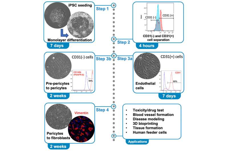
Graphical abstract. Credit: STAR Protocols (2023). DOI: 10.1016/j.xpro.2023.102292
Researchers at NIH's National Eye Institute have published a detailed protocol for making three cell types that are key components to form blood vessels and capillaries. The technique enables researchers to make tissues for study and potentially for future treatments.
"This protocol generates vascular endothelial cells, pericytes, and fibroblasts from human induced pluripotent stem cells called iPSCs," said Tea Soon Park, Ph.D., NEI Ocular and Stem Cell Translational Research Section, and lead author of the protocol.
IPSCs are made in a lab, starting with skin or blood cells. They can then be modified to create nearly any other cell type of the body. "Since all three cell types in this protocol are major parts of vascular structure, it will be a useful tool to study a variety of diseases including eye disease," said Park.
Credit: NIH, National Eye Institute (NEI)
The five-step protocol, from iPSCs to fibroblasts, takes about four weeks. It includes seeding of iPSCs, seven days of monolayer differentiation, and cell separation based on CD31 expression. CD31 is a cell-surface protein unique to endothelial cells. At the end of step 3, endothelial and pericyte populations are separated. Fibroblasts can then be further differentiated from pericytes.
Park and colleagues are now working on a method to combine the three patient-derived cell types using bioprinting, which distributes and orients cells in a 3D space resembling the tissue microenvironment.
The paper is published in the journal STAR Protocols.


Post comments