
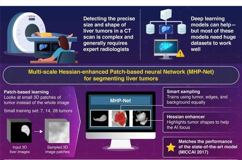
Researchers from Science Tokyo develop a Multi-scale Hessian-enhanced Patch-based Neural Network Model for Segmentation of Liver Tumor from CT Scans. Credit: Institute of Science Tokyo, Japan
Liver cancer is the sixth most common cancer globally and a leading cause of cancer-related deaths. Accurate segmentation of liver tumors is a crucial step for the management of the disease, but manual segmentation by radiologists is labor-intensive and often results in variations based on expertise.
Artificial intelligence (AI)-based tumor segmentation models have revolutionized tumor assessment in medical imaging—using deep convolutional neural networks, they identify and outline the exact shape, size, and location of a tumor in a medical scan image. But their efficacy comes with a heavy dependence on large volumes of data (typically ranging from 1, 000 to 10, 000 cases). This requirement for big data is a major barrier in medical AI.
To overcome this barrier, a team of researchers led by Professor Kenji Suzuki and a Ph.D. student, Yuqiao Yang, from the Biomedical AI Research Unit of Institute of Science Tokyo (Science Tokyo), Japan, has developed a groundbreaking AI model that can accurately segment liver tumors from computed tomography (CT) scans—even when trained using extremely small datasets—surpassing the performance of current state-of-the-art systems. Their study is published in the journal IEEE Access.
At the heart of this innovation is a novel architecture called the multi-scale Hessian-enhanced patch-based neural network (MHP-Net). MHP-Net works by breaking medical images into small 3D image patches—so the AI can focus on one part at a time rather than the entire image. It then pairs each patch from the original CT image with a corresponding enhanced version, achieved through a technique called Hessian filtering. Hessian filtering helps highlight spherical objects such as tumors in the image.
The result is a high-resolution tumor segmentation map that accurately delineates liver tumors from contrast-enhanced CT scans. To evaluate the model's performance, the team used the "Dice similarity score, " which compares how well the predicted segmentation matches the ground truth (usually annotated by expert radiologists) on a scale of 0 to 1.
"Despite a limited training set of 7, 14, and 28 tumors, we achieved high performance dice scores of 0.691, 0.709, and 0.719, respectively, " notes Suzuki. "With these scores, our model surpasses major established models such as U-Net, Res U-Net, and HDense-U-Net."
Apart from its promising performance, the lightweight architecture of the model allows for fast training (under 10 minutes) and real-time inference (~4 seconds per patient), making it highly suitable for use even in clinical settings with limited computational resources.
"This is just a start in the field of small-data AI, where meaningful and clinically relevant deep learning models can be built from limited datasets, " says Suzuki. "MHP-Net's success can inspire small-data AI solutions in other areas of medical imaging as well, such as the detection of rare cancers."
The study shows the potential of small-data AI in medical image analysis. By lowering the threshold for the data required for training, MHP-Net democratizes the use of AI in medical image analysis, especially in under-resourced hospitals and clinics with limited access to data. In the future, the researchers plan to explore broader applications of small-data AI models—enabling scalable, cost-effective, and versatile deployment of AI in health care worldwide.
More information: Yuqiao Yang et al, Patch-Based Deep-Learning Model With Limited Training Dataset for Liver Tumor Segmentation in Contrast-Enhanced Hepatic Computed Tomography, IEEE Access (2025). DOI: 10.1109/ACCESS.2025.3570728 Journal information: IEEE Access


Post comments