
by Fabienne Landry, McGill University Health Centre
Credit: Cell (2024). DOI: 10.1016/j.cell.2024.06.023
Posterior fossa group A (PFA) ependymoma are rare, treatment-resistant pediatric tumors of the central nervous system that originate in the brain and spinal cord. They have the highest recurrence rate and poorest prognosis of all childhood cancers due to the lack of effective treatment.
Hope is on the horizon now that an international research team led by scientists at Baylor College of Medicine in Texas, U.S., and the Research Institute of the McGill University Health Center (RI-MUHC) in Montreal, Canada, have identified unique 3-dimensional features called TULIPs in the genome of PFA ependymoma that could eventually be targeted in the development of more effective therapies.
The findings are published in the journal Cell.
"PFA ependymomas are lethal. One of the reasons behind the little progress toward the development of effective treatments for these tumors is that the majority of PFAs lack clear genetic mutations driving tumor growth.
"Without a clear genetic target against which we could design specific therapies, we investigated another aspect of the tumor, how the DNA is packed inside the nucleus of the cell," says senior and lead author, Marco Gallo, Associate Professor of pediatrics, hematology-oncology at Baylor and Texas Children's Hospital.
"Our work was driven by a simple fact: PFA ependymoma are usually diagnosed in very young children, and have no efficient treatment. Radiation therapy, the only treatment currently available, is not effective and causes serious developmental and cognitive issues. That's a reality we hope to change," says Canada Research Chair in Pediatric Oncology Dr. Nada Jabado, co-senior author of the study, a Senior Scientist in the Child Health and Human Development Program at the RI-MUHC and a Pediatric Hemato-Oncologist at the Montreal Children's Hospital of the MUHC.
Uncovering the 3D configuration of tumor cells' genomes
Every cell in the body has about 6.6 feet (2 meters) of linear DNA that is stored in its nucleus in a manner that enables the cell to easily access the genes it uses most often and set aside those less used. This would be like organizing the closet with the clothes most frequently used at the front and those rarely worn on the back.
To fit in the tiny nucleus, the long DNA molecules are folded, twisted, looped, which results in specific 3D conformations, some tighter, some more relaxed, that can ultimately help the cell express the genes needed to do its job.
In this study, the researchers looked closely at what we might call the "geography" of the PFA ependymoma cell genome (the entire set of DNA instructions found in the cell).
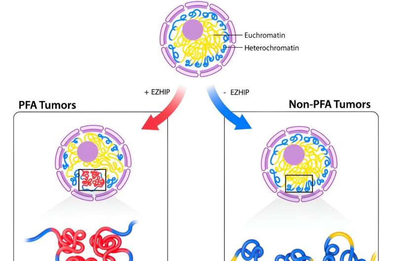
"We investigated the unique ways PFAs cells organize their DNA in 3D, by orchestrating strong interactions between regions of the genome that are normally very far apart. We discovered specific regions that are not present in other types of pediatric brain cancer and that recur at predictable genomic locations.
"We named them TULIPs, for Type B Ultra-Long Interactions in PFAs," says Dr. Michael D Taylor, co-senior author of the study and Professor of pediatrics, hematology—oncology and neurosurgery at Baylor and Texas Children's. He also is the Cyvia and Melvyn Wolff Chair of Pediatric Neuro-Oncology at Texas Children's Cancer and Hematology Center.
The researchers used the Hi-C technology to profile the 3D architectures of the entire genomes of PFA tumors and compared them with those of a large cohort of samples from different tumor types and nonmalignant tissues. In the process, TULIPs appeared as specific regions of very tightly compacted, thus hard-to-access, DNA, a sign that the cell may not use the genes in that region often.
"TULIPs also tend to interact with each other over very long distances. A TULIP might be at the end of a chromosome and another TULIP at the other end of the same chromosome, and they find their way to interact with each other with surprising strength," explains Prof. Gallo.
"TULIPs on different chromosomes can also converge and strongly interact with each other. We also found that regions outside TULIPs appear more relaxed overall. This is important because TULIPs are linked to the cell's function."
A potentially actionable chemical tag
According to the study findings, TULIPs carry a methyl group on histone H3K9, a protein associated with DNA that can act as a chemical tag. Indeed, when the research team inhibited the tagging of H3K9 in PFA patient-derived cultures, they witnessed weaker interactions between TULIPs and impaired PFA cell survival. These observations suggest that TULIP interactions are important for PFA cell viability, opening up new potential targets for treatment.
"We believe that TULIPs are ephemeral structures present at an early stage of cancer development in progenitor cells—cells that descend from stem cells and precede the creation of mature cells, very early in life. However, more research is needed to understand the mechanism by which TULIPs arise and mediate cancer cells behavior," says Dr. Jabado, who is also a professor in the Department of Pediatrics at McGill University.
"By further investigating this mechanism, we may be able to explore treatment strategies to remove them to promote tumor elimination."
More information: Michael J. Johnston et al, TULIPs decorate the three-dimensional genome of PFA ependymoma, Cell (2024). DOI: 10.1016/j.cell.2024.06.023
Journal information: Cell
Provided by McGill University Health Centre


Post comments