
Credit:Gastric cancer-derived exosomal miR-519a-3p promotes liver metastasis by inducing intrahepatic M2-like macrophage-mediated angiogenesis
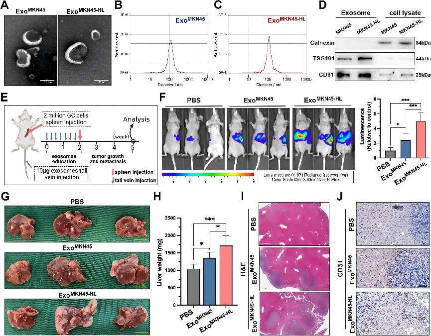
GC-derived exosomes promote liver metastasis. A. Representative images of exosomes from MKN45 and MKN45-HL cells at TEM, Scale bar = 100 nm. B, C. Purified MKN45 and MKN45-HL exosomes were analyzed by NanoSight. D. Western blot analysis of exosome markers (TSG101 and CD81) in exosomes and lysates from MKN45 and MKN45-HL cells. E. Schematic representation of the establishment process of the exosome-educated GC-LM model. F. Representative in vivo imaging system (IVIS) results of mice injected with luciferase-labeled MKN45 cells into the spleen after educated with PBS, MKN45 exosomes, or MIN45-HL exosomes, respectively. Results of quantified values of bioluminescence imaging signals are expressed as mean ± standard deviation (n = 6). G-I. Effect of GC-derived exosomes on liver metastasis in mice. The representative photographs of liver metastasis, liver weight, and H&E staining were shown. The scale bars are 1.0 cm and 500 μm, respectively. J. Representative images of CD31 immunohistochemical staining of LM tissues from mice with exosome education, Scale bar = 100 μm. Data are shown as mean ± standard deviation of 3 independent experiments, and statistical significance was determined using one-way ANOVA test (*P < 0.05, **P < 0.01, ***P < 0.001)
Liver metastasis (LM) is one of the most common types of metastasis in gastric cancer (GC) patients, significantly impacting their survival rate and quality of life. The occurrence of gastric cancer liver metastasis involves complex biological processes, including tumor cell infiltration, angiogenesis, and immune escape. Exosomes are extracellular vesicles with a diameter of approximately 30-150 nanometers, widely present in body fluids. They are capable of carrying proteins, RNA, DNA, and other biomolecules, and participate in intercellular communication and material exchange. Studies have shown that tumor cells can promote growth, invasion, and metastasis by releasing exosomes that alter the behavior of surrounding cells. miRNAs in exosomes play a critical regulatory role, transmitting information between cells and altering the gene expression profiles of recipient cells, thereby affecting their function. However, the specific mechanisms of exosomal miRNAs in tumor metastasis require further investigation.
Recent research by Qiu et al. revealed the regulatory mechanism of miR-519a-3p in exosomes derived from gastric cancer (GC) cells in the context of gastric cancer liver metastasis (GC-LM). Exosomes were extracted and identified using differential centrifugation, transmission electron microscopy (TEM), and nanoparticle tracking analysis (NTA). The role of GC-derived exosomes and exo-miR-519a-3p in angiogenesis and GC-LM was investigated through tube formation assays, aortic ring assays, and an exosome-educated GC-LM model. Various techniques such as dual-luciferase reporter assays, qRT-PCR, Western blotting, ELISA, flow cytometry, and immunofluorescence were employed to explore the regulatory mechanisms of exo-miR-519a-3p in GC-LM.
The results demonstrated that the expression of miR-519a-3p in the serum exosomes of GC-LM patients was significantly higher than in patients without liver metastasis. High expression of exo-miR-519a-3p was associated with poor prognosis. GC-derived exosomes predominantly accumulated in the liver and were internalized by hepatic macrophages. The study further showed that exo-miR-519a-3p could target DUSP2 to activate the MAPK/ERK pathway, inducing M2 polarization of macrophages. These M2-polarized macrophages then promoted angiogenesis and the formation of a pre-metastatic niche in the liver, accelerating GC-LM.
In in vivo experiments, GC-derived exosomes significantly increased the fluorescence intensity in the livers of mice, particularly in those educated with exosomes from high liver metastasis potential MKN45-HL cells. In vitro experiments demonstrated that MKN45-HL exosomes significantly increased the expression of M2 macrophage markers and decreased the expression of M1 markers in PMA-treated THP-1 cells. In tube formation and aortic ring assays, THP-1 cells treated with MKN45-HL exosomes significantly increased the number of tubular structures formed by HUVECs and the number of sprouts in mouse aortic rings.
In summary, this study indicates that high expression of exo-miR-519a-3p could serve as a potential diagnostic biomarker for GC-LM, as its serum levels are closely associated with the prognosis of GC-LM patients. Therefore, targeting exo-miR-519a-3p could be a promising therapeutic strategy for GC-LM.
This study is the first to identify the critical role of miR-519a-3p in gastric cancer liver metastasis (GC-LM) and to elucidate the mechanism by which it promotes angiogenesis and liver metastasis through the induction of M2 polarization of hepatic macrophages. This finding provides a theoretical basis and experimental support for the development of new anti-tumor drugs and therapeutic methods. Additionally, the study employs multi-level experimental approaches, including in vitro cell experiments and in vivo animal models, to comprehensively validate the function and molecular mechanisms of miR-519a-3p. Such multi-level validation enhances the credibility and scientific rigor of the research findings.
However, many issues and challenges still need to be addressed in future research. The significant variations in the size, content, and composition of exosomes could affect their function and delivery efficiency in the tumor microenvironment due to this heterogeneity. Secondly, limited research exists on the delivery mechanisms of exosomes in vivo, and the distribution of exosomes in different tissues and organs, as well as their stability and persistence in the body, has not been thoroughly elucidated. Moreover, the study lacks an investigation into other potential signaling pathways and molecules that may be involved in M2 macrophage polarization, which might result in an incomplete understanding of this process.
Future research can focus on exploring the heterogeneity of exosomes and their functional differences, optimizing delivery systems, and utilizing multi-omics technologies and gene editing techniques to systematically analyze the regulatory mechanisms of M2 macrophage polarization. This approach may help identify new therapeutic targets. By advancing these research methodologies and focal areas, significant progress can be made in addressing the current study's limitations and challenges. This, in turn, will further the development of research on gastric cancer liver metastasis and exosomes, providing more reliable scientific evidence and technical support for clinical applications.
Qiu, Shengkui, et al. "Gastric cancer-derived exosomal miR-519a-3p promotes liver metastasis by inducing intrahepatic M2-like macrophage-mediated angiogenesis." Journal of Experimental & Clinical Cancer Research 41.1 (2022): 296.


Post comments