
Credit: JACS Au (2025). DOI: 10.1021/jacsau.5c00151
Tumor cells have higher metabolic activity compared to healthy tissue and consequently consume a lot of glucose. Positron Emission Tomography (PET)—the current gold standard diagnostic technique—exploits this property.
Clinicians inject patients with radioactive tracers such as 18F-fluoro 2-deoxy glucose (18F-FDG), which accumulate at the tumor site and help pinpoint it. However, PET is expensive, and poses the risk of radiation accumulation in the case of repeated scans.
Researchers from the Department of Bioengineering, Indian Institute of Science (IISc), have now developed a biocompatible small molecule that can be used as a minimally invasive contrast agent for photoacoustic (PA) tomography. Their study has been published in JACS Au.
In this method, a near-infrared (NIR) laser beam is shined on light-absorbing molecules (chromophores) sent to the target region, which then expand, creating a pressure change. The change can be picked up as an auditory signal, and analyzing these signals allows scientists to construct 3D images of the target region. The method is particularly useful for pinpointing superficial tumors.
"You are able to use a more cost-effective technique, cheaper than both PET and Magnetic Resonance Imaging (MRI), and get the same information, " says Sanhita Sinharay, assistant professor at BE and corresponding author of the study.
Currently, photoacoustic imaging in a clinical setting only makes use of natural chromophores like hemoglobin, which are already present in the human body. Its oxygenated and deoxygenated states have different optoacoustic signatures that act as useful biomarkers of abnormal metabolic processes.
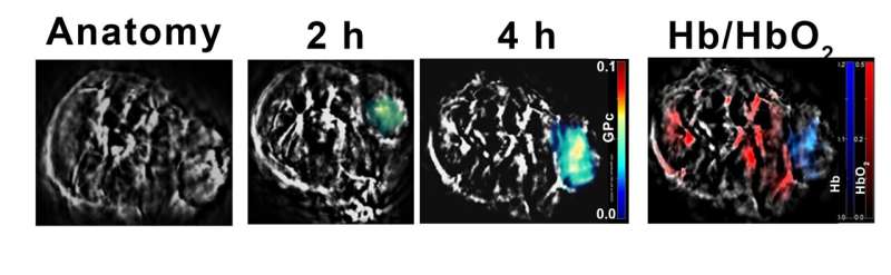
MSOT image of a tumor-bearing mouse demonstrating maximal uptake of GPc in 4h, overlayed on the anatomy and an Hb/HbO2 map in the same tumor, highlighting the low oxygen gradient in this tumor. Credit: Pooja Patkulkar
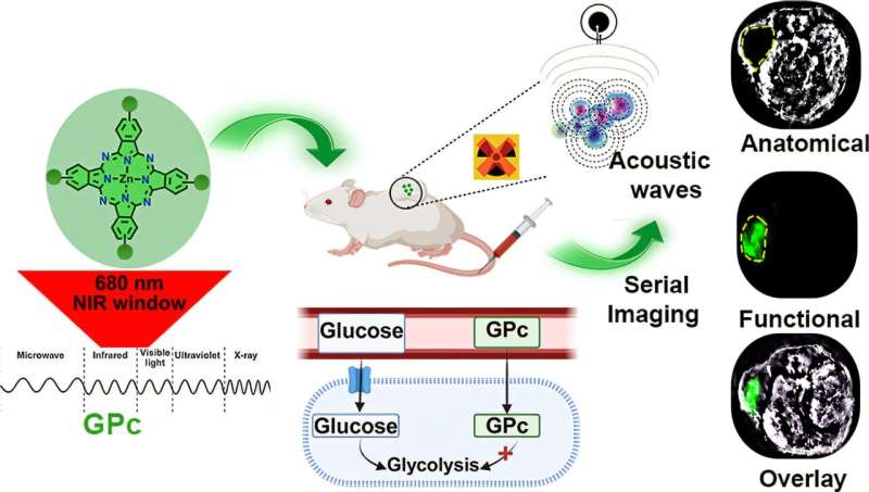
In the new study, the IISc team designed an external molecule—not typically found in human cells—to provide better sensitivity and contrast, which makes it easier to differentiate the target region from normal tissues. The molecule, referred to as GPc, consists of four glucose units conjugated to a scaffold made of zinc-phthalocyanine.
Phthalocyanines are chemicals that, when excited in the NIR window, dissipate energy primarily through a nonradiative process and therefore are well suited for photoacoustic imaging.
While designing GPc, the researchers expected that adding four glucose units to it would improve the cellular uptake of the molecule. Adding glucose, with its four water-loving hydroxyl groups, also increased the molecule's solubility.
"One of the major milestones for us was the mechanistic evaluation of the probe with the seahorse assay, " explains Pooja Patkulkar, Ph.D. student at BE and first author of the study.
"We wanted to see whether the molecule we made was being taken up by the glucose transporters, and what its fate was after uptake. The aim was to compare its behavior with 18F-FDG, and see whether it is a glucose antagonist or agonist."
An agonist would compete with glucose and be metabolized by the cell, and thus would be an ineffective contrast agent. In order to understand the exact nature of the new agent, the team set up a competitive chemical assay between GPc and glucose.
They saw that GPc entered human cells easily, was not metabolized, and its uptake was not facilitated by GLUT1 transporters that enable the uptake of glucose. The authors, however, suggest that more studies are needed to fully evaluate GPc's function once it enters the cells.
"The strength of our work was proving to some extent the behavior of the probe inside cells and validating its entry into the tumor core which exhibits a low gradient of molecular oxygen supply, " says Sinharay.
"Once it enters the tumor, how is it functioning? How close is the behavior to 18F-FDG? Even though the structure and molecular weight are different, their functionality is very close. I think that was really surprising."
The authors note that such molecules can greatly help visualize tumor sites with high metabolic activity non-invasively. The use of such molecules can also circumvent challenges with PET, while providing similar functionality at a lower cost for pinpointing superficial tumors.
More information: Pooja A. Patkulkar et al, In Vivo Visualization of Tumor Metabolic Activity with a Tetra Glucose-Conjugated Zinc-Phthalocyanine Photoacoustic Contrast, JACS Au (2025). DOI: 10.1021/jacsau.5c00151 Journal information: JACS Au


Post comments