
by Bill Snyder, Vanderbilt University
Credit: Gastroenterology (2023). DOI: 10.1053/j.gastro.2023.04.038
Fibroblast cells play key roles in the repair of damaged tissue and in pathological scarring. Now, researchers at Vanderbilt University Medical Center have uncovered evidence of their direct involvement in the development of gastric cancer.
These findings, published in the journal Gastroenterology, could lead to novel interventions to prevent cancer of the stomach, the third leading cause of cancer deaths worldwide after lung and colorectal malignancies.
Until recently it has been difficult to identify which cells are involved in the steps leading to gastric cancer, notably the development of precancerous stomach lesions called metaplasia, and later, dysplasia, the appearance of abnormal cells.
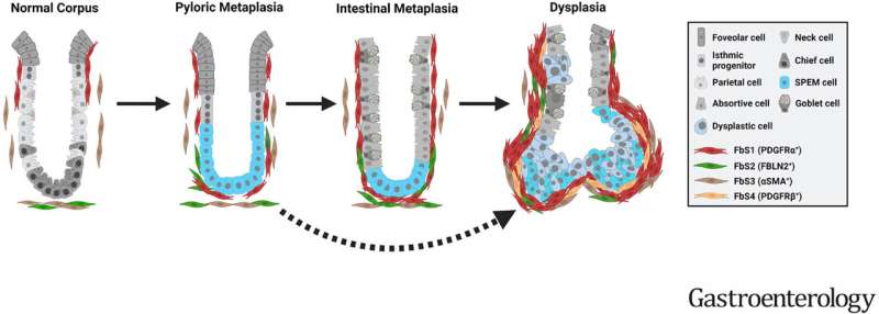
"Our studies demonstrated that particular subsets of fibroblasts present in the stomachs of patients with metaplasia and cancer can promote transition of precancerous metaplasia towards dysplasia," said James Goldenring, MD, Ph.D., the Paul W. Sanger Professor of Surgery and vice chair for research in the Section of Surgical Sciences.
In 1999 Goldenring and his colleagues were the first to describe spasmolytic polypeptide-expressing metaplasia (SPEM), a lineage of metaplastic cells in the stomach that develop from normal protein-secreting cells after damage to the stomach lining. Chronic persistence of these cells can lead to the development of gastric cancer.
For several years they have been using metaplastic and dysplastic gastroids, three-dimensional cultures of epithelial cells isolated from the stomach lining of mice and humans, to identify the characteristics of precancerous stem cell populations.
In the latest study, the researchers isolated four distinct subsets of fibroblasts from the stroma, the tissue surrounding the stomach lining, in healthy people and those with gastric cancer.
One subset of fibroblasts was expanded in patients with metaplasia and cancer compared to healthy controls. When cocultured with SPEM gastroids, these metaplasia- or cancer-derived fibroblasts induced disordered growth, loss of metaplastic cell markers, and an increase in dysplastic cell markers, indicative of the transition of metaplastic cells towards cancer.
Culturing metaplastic gastroids with conditioned media from the laboratory dishes in which these fibroblasts were grown also promoted the dysplastic transition, suggesting that dysplastic progression can be accelerated by factors secreted by the fibroblasts in proximity to metaplastic epithelial cells in the stomach lining.
These findings indicate that SPEM lineage cells under the influence of altered fibroblast populations may evolve to dysplasia without going through intermediate steps.
While further study is needed, targeting this subset of fibroblasts and the factors they secrete may be an effective way to block the progression from precancerous stomach lesions to gastric cancer, the researchers concluded.
More information: Su-Hyung Lee et al, Apposition of Fibroblasts With Metaplastic Gastric Cells Promotes Dysplastic Transition, Gastroenterology (2023). DOI: 10.1053/j.gastro.2023.04.038
Journal information: Gastroenterology
Provided by Vanderbilt University


Post comments