
Mechanistic Insights and Therapeutic Potential of Jmjd4 in Regulating Cardiomyocyte Metabolism in Dilated Cardiomyopathy
Deleting Jmjd4 in adult cardiomyocytes causes progressive dilated cardiomyopathy and heart failure.
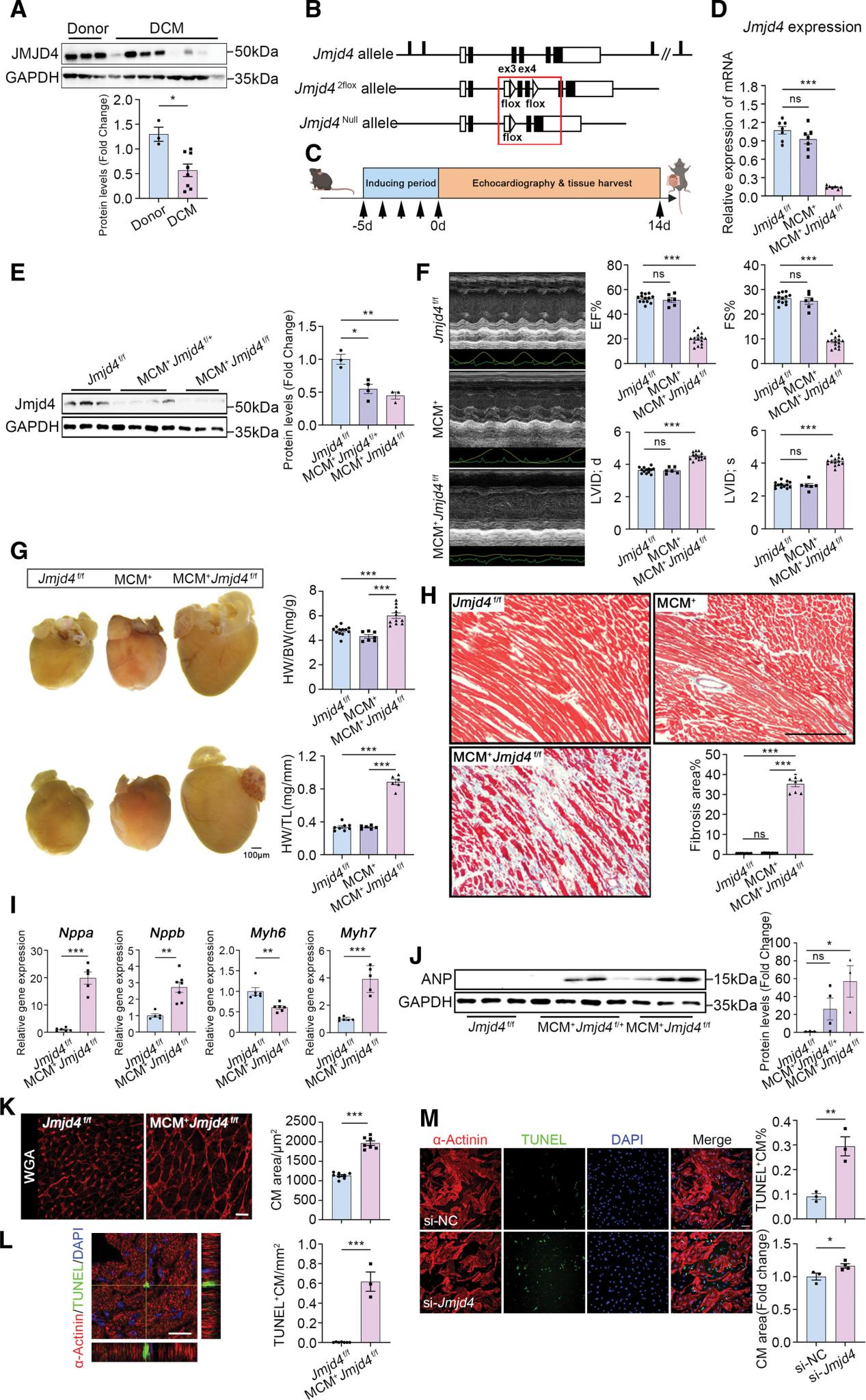
A, Western blot and quantification of JMJD4 protein levels in the extract of heart tissues from human patients with DCM and healthy donors (n=3 healthy donors for donor group, n=8 patients for DCM group). B, Strategy of generating a floxed allele of Jmjd4 in mouse. Maps of the WT Jmjd4 locus, the floxed allele, and the excised allele are shown. Exons are shown in bars. C, Schematic of tamoxifen-induced Jmjd4 knockout in the adult MCM; Jmjd4+f/f mouse hearts. D, Quantitative real-time polymerase chain reaction analysis of Jmjd4 mRNA levels in the heart tissue of Jmjd4f/f, MCM, and MCM; Jmjd4++f/f mice (n=7 animals per group). Data from MCM; Jmjd4+f/f mice in D through L were acquired at 14 days after tamoxifen induction. E, Western blot and quantification of Jmjd4 protein levels in the extract of heart samples from Jmjd4f/f, MCM; Jmjd4+f/+, and MCM; Jmjd4+f/f mice (n=3 animals/Jmjd4f/f, 4 animals/MCM; Jmjd4+f/+, 3 animals/MCM; Jmjd4+f/f). F, Representative M-mode echocardiographic images and corresponding measurements of ejection fraction (EF), fractional shortening (FS), and internal dimension of the left ventricle in diastole (LVID;d) and systole (LVID;s) of Jmjd4f/f, MCM, and MCM; Jmjd4++f/f mice (n=13 animals/Jmjd4f/f, 6 animals/MCM, 15 animals/MCM; Jmjd4++f/f). G, Gross morphology of the hearts (scale bar, 100 μm), ratios of heart weight to body weight (HW/BW), and bi-atrial heart weight to tibia length (HW/TL) in Jmjd4f/f, MCM and MCM; Jmjd4++f/f mice at 3 months of age (n=12 animals/Jmjd4f/f, 7 animals/MCM, 11 animals/MCM; Jmjd4++f/f). H, Representative Masson trichrome staining of cross-sections (scale bars, 100 μm) and quantification of fibrotic areas of the hearts of Jmjd4f/f, MCM, and MCM; Jmjd4++f/f mice (n=8 animals per group). I, Quantitative real-time polymerase chain reaction analysis of mRNA levels of Nppa, Nppb, Myh6, and Myh7 genes from the heart tissue of Jmjd4f/f and MCM; Jmjd4+f/f mice (n=6 animals/Jmjd4f/f, 5 animals/MCM; Jmjd4+f/f). J, Western blot and quantification of atrial natriuretic peptide (ANP) protein levels from the heart apex tissue of Jmjd4f/f, MCM; Jmjd4+f/+, and MCM; Jmjd4+f/f mice (n=3 animals/Jmjd4f/f, 4 animals/MCM; Jmjd4+f/+, 3 animals/MCM; Jmjd4+f/f). K, Representative images of wheat germ agglutinin (WGA) staining and quantification of mean cardiomyocyte cross-section area of ventricular cardiomyocytes (CM) from hearts of Jmjd4f/f and MCM; Jmjd4+f/f mice (n=7 animals per group). L, Representative images of terminal deoxynucleotidyl transferase dUTP nick end labeling (TUNEL) and α-actinin staining and quantification of cell death of cardiomyocytes in Jmjd4f/f and MCM; Jmjd4+f/f hearts (n=7 animals/Jmjd4f/f, 3 animals/MCM; Jmjd4+f/f). M, Representative images of fluorescence staining of TUNEL and α-actinin (scale bar, 100 μm) in neonatal rat ventricular cardiomyocytes 96 hours after being transfected with small interfering negative control (si-NC) or si-Jmjd4, and quantified as the percentage of TUNEL-positive cells in α-actinin–expressing cardiomyocytes (n=3 wells per group). One-way ANOVA with Bonferroni correction was performed (D–H, J). Two-tailed unpaired Student t test was performed (A, I, K–M). All in vitro experiments were assessed 3 times independently. All quantitative data are expressed as mean±SEM, *P<0.05, **P<0.01, ***P<0.001. DAPI indicates 4’,6-diamidino-2-phenylindole; DCM, dilated cardiomyopathy; and ns, not significant.
From:Jmjd4 facilitates Pkm2 degradation in cardiomyocytes and is protective against dilated cardiomyopathy.
Dilated cardiomyopathy (DCM) is a severe cardiovascular disease characterized by ventricular dilation and impaired cardiac contraction, eventually leading to heart failure. Previous studies have suggested that abnormal responses to metabolic stress play a crucial role in the pathogenesis of DCM, but the specific molecular mechanisms remain unclear. The JMJD family proteins, initially identified as histone deacetylases, have now been found to play diverse roles in various cardiovascular diseases. However, whether JMJD family members are involved in DCM remains unknown. In a recent study, Tang et al. investigated the role of Jmjd4 in DCM by analyzing cardiac tissues from DCM patients and mouse models, exploring how Jmjd4 regulates cardiomyocyte metabolism and assessing its potential as a therapeutic target.
Jmjd4 Expression and Cardiac Function
Western blot analysis of cardiac tissues from DCM patients revealed that Jmjd4 expression was significantly lower compared to healthy controls. To further understand Jmjd4's role in DCM progression, the researchers conditionally knocked out Jmjd4 in mouse cardiomyocytes. These mice displayed typical DCM symptoms, including cardiac enlargement and significantly reduced contractile function. Using the Myh6-CreERT2 system, Jmjd4 was conditionally knocked out in mouse cardiomyocytes, resulting in significant metabolic abnormalities and myocardial hypertrophy. The study observed that the expression of Nppa and Nppb genes was significantly upregulated, while the expression of the Myh6 gene was downregulated in cardiomyocytes. These findings were confirmed through siRNA-mediated Jmjd4 gene knockdown experiments.
Molecular Mechanisms and Metabolic Pathways
RNA sequencing and metabolomic analysis indicated that the loss of Jmjd4 led to the downregulation of various metabolism-related genes in cardiomyocytes, particularly those associated with mitochondrial function and glycolysis. Additionally, Jmjd4 deficiency caused a significant reduction in mitochondrial membrane potential and decreased mitochondrial respiration rates. Mass spectrometry and co-immunoprecipitation experiments demonstrated that Jmjd4 directly interacts with Pkm2. Further experiments revealed that Jmjd4 hydroxylates Pkm2 at the K66 site, promoting its degradation through the chaperone-mediated autophagy pathway. This finding underscores the critical role of Jmjd4 in regulating cardiomyocyte metabolism.
Therapeutic Potential of Targeting Pkm2
The study also explored the therapeutic potential of targeting Pkm2. The use of the Pkm2 agonist TEPP-46 showed significant therapeutic effects in both the Jmjd4 deficiency-induced DCM model and the pressure overload-induced heart failure model. This suggests that activating the underperforming Pkm2 can alleviate cardiac metabolic dysfunction induced by various stresses.
Methodological Strengths and Limitations
This research presents a novel approach, revealing that Jmjd4 promotes the degradation of Pkm2 through its interaction with heat shock protein Hsp70. This mechanism offers new insights into the metabolic regulation of DCM and provides potential avenues for developing new therapeutic strategies. Unlike traditional research methods that focus on the function of a single gene or protein, this study employed whole-genome and metabolomic analyses, allowing for a more comprehensive understanding of Jmjd4's impact on cardiomyocyte metabolism. By using a conditional gene knockout mouse model, the researchers specifically knocked out Jmjd4 in adult cardiomyocytes, avoiding developmental abnormalities associated with embryonic knockout. This method more accurately reflects the function of Jmjd4 in adult cardiomyocytes.
However, there are limitations to this study. The sample size was relatively small, particularly in the patient samples, which included only a few DCM patients and healthy controls. This limitation may affect the generalizability of the results. Moreover, some cellular-level changes observed in vitro may not fully reflect the complex physiological environment in vivo. Additionally, other potential substrates of Jmjd4 and its interactions with other molecular pathways have not been fully elucidated.
Future Directions
Future research should expand the sample size, delve deeper into the mechanisms, and assess the feasibility and safety of clinical applications to provide a more reliable foundation for DCM treatment. By further exploring the multiple roles of Jmjd4 in cardiomyocyte metabolism, the specific regulatory mechanisms of Pkm2, and its clinical translation as a therapeutic target, this research could significantly advance the development and application in this field. These studies not only contribute to a better understanding of the pathophysiological mechanisms of DCM but also have the potential to foster new therapeutic strategies and technologies, opening up extensive opportunities for industrial development.
Reference:
Tang, Yansong, et al. "Jmjd4 facilitates Pkm2 degradation in cardiomyocytes and is protective against dilated cardiomyopathy." Circulation 147.22 (2023): 1684-1704.


Post comments