

Introduction
Eyelid malignancies, including sebaceous carcinoma (SC) and squamous cell carcinoma (SCC), present significant diagnostic challenges with important clinical implications. These tumors can invade locally, metastasize, and compromise both ocular function and patient survival. Histopathologic differentiation between SC and SCC is notoriously difficult due to overlapping morphologic features—such as poor differentiation and similar cellular architecture—that often confound diagnosis.
Sebaceous carcinoma, in particular, may mimic SCC or basal cell carcinoma because of its frequent squamous epithelialization and loss of typical sebaceous differentiation. Reliance solely on hematoxylin and eosin (H&E) staining increases the risk of misdiagnosis, which can delay appropriate treatment or lead to inappropriate therapeutic decisions, ultimately worsening patient outcomes. This diagnostic dilemma underscores the urgent need for enhanced, reliable, and standardized diagnostic approaches.
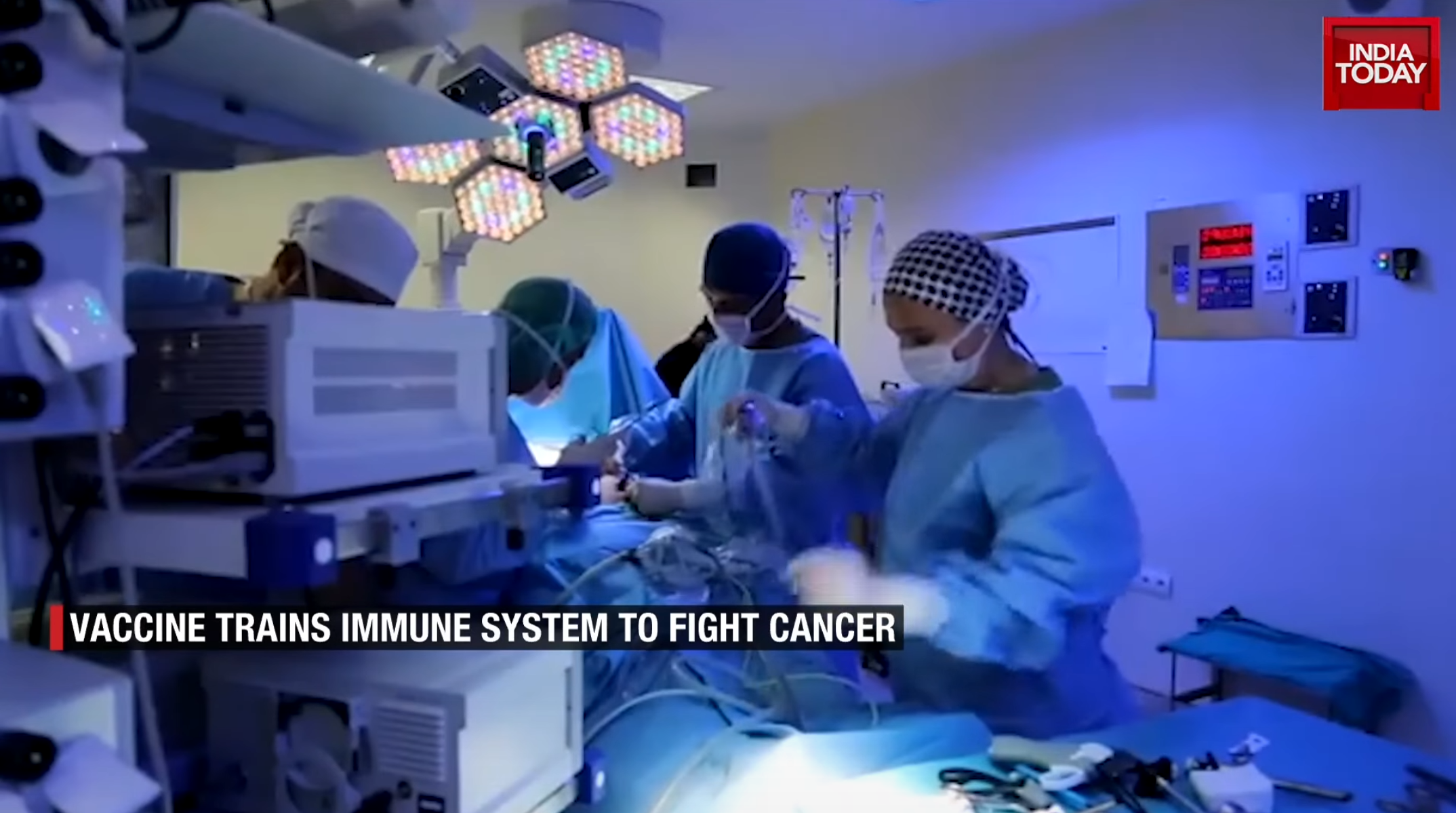
Recent advances in artificial intelligence (AI) have introduced a promising solution. By leveraging deep learning and large-scale histopathologic image analysis, AI systems can improve accuracy, reproducibility, and efficiency in distinguishing SC from SCC, particularly in challenging or poorly differentiated cases.1,2,3,4
Artificial Intelligence as a Diagnostic Partner
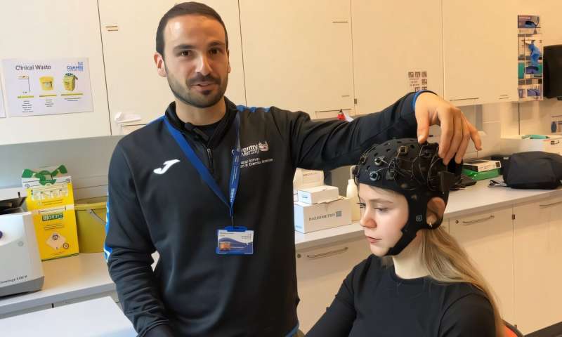
AI is increasingly transforming histopathology through the application of machine learning, deep learning, and convolutional neural networks (CNNs). These systems are trained on extensive datasets of digitized whole slide images (WSIs), following a pipeline of image acquisition, preprocessing such as tissue segmentation and tile extraction, feature extraction, and classification.
AI models can detect subtle morphologic and textural patterns beyond the limits of human perception, reducing interobserver variability and enhancing diagnostic consistency. Furthermore, AI-driven triage can highlight slides that warrant urgent review, optimizing workflow and partially alleviating the global shortage of pathologists.
Recent innovations such as weakly supervised learning and vision transformers allow for more comprehensive slide-level analysis by integrating patch-based features into a global diagnostic context. These methods not only improve tumor classification but also characterize the surrounding microenvironment, offering deeper insights into tumor biology. AI tools also streamline labor-intensive tasks such as mitotic figure quantification, immunohistochemical analysis, and biomarker detection, supporting both diagnostic and predictive aspects of precision oncology. 5,6,7
AI in Differentiating Sebaceous vs Squamous Cell Carcinoma
Deep learning algorithms have shown particular promise in distinguishing SC from SCC of the eyelid—two entities that often present with overlapping histopathologic features. Trained on expert-annotated WSIs, these models can recognize nuanced differences such as the lipid-rich cytoplasmic texture characteristic of sebaceous cells, the distinct nuclear contours, and the specific chromatin distribution and keratinization patterns typical of squamous carcinomas.
Some research groups have advanced this further by integrating immunohistochemistry data into multimodal AI models, allowing the systems to correlate morphologic features with molecular biomarkers. This approach has improved sensitivity, specificity, and overall diagnostic accuracy, with some models achieving performance comparable to, or exceeding, that of experienced pathologists. AI-aided diagnosis is particularly valuable in borderline or poorly differentiated cases, where histopathologic interpretation is most challenging. Visualization tools such as heatmaps and attention maps further enhance transparency by showing pathologists which regions and features informed the AI’s decision, fostering greater trust in AI-assisted workflows. 8,9,10
Conclusion
The integration of AI into histopathologic practice represents a paradigm shift in ocular oncology diagnostics. By quantitatively analyzing complex morphologic and molecular patterns, AI offers a powerful adjunct to the pathologist’s expertise, improving diagnostic accuracy, consistency, and efficiency.
As AI systems continue to evolve—incorporating larger, more diverse datasets and multimodal information—they hold the potential to become indispensable tools in differentiating sebaceous carcinoma from squamous cell carcinoma of the eyelid, ultimately improving patient outcomes through earlier and more precise treatment decisions.
Reference:
1.Yunoki T, Miyakoshi A, Hayashi A. Clinicopathologic Features of Eyelid Sebaceous Gland Carcinoma Requiring Immunohistochemical Diagnosis. Ocul Oncol Pathol. 2024 Sep;10(3):131-138. doi: 10.1159/000538537. Epub 2024 Apr 29. PMID: 39224525; PMCID: PMC11368396.
2.Geng J, Zhang K, Dong L, Hui S, Zhang Q, Li Z, Zhang R, Jiang X, Wang M, Sun S, Zhang H, Yang Y, Yang X, Piao Y, Li D. AI assistance enhances histopathologic distinction between sebaceous and squamous cell carcinoma of the eyelid. NPJ Digit Med. 2025 Jul 4;8(1):406. doi: 10.1038/s41746-025-01775-z. PMID: 40615496; PMCID: PMC12227637.
3.Choi S, Kim S. Artificial Intelligence in the Pathology of Gastric Cancer. J Gastric Cancer. 2023 Jul;23(3):410-427. https://doi.org/10.5230/jgc.2023.23.e25
4.Semerci ZM, Toru HS, Çobankent Aytekin E, Tercanlı H, Chiorean DM, Albayrak Y, Cotoi OS. The Role of Artificial Intelligence in Early Diagnosis and Molecular Classification of Head and Neck Skin Cancers: A Multidisciplinary Approach. Diagnostics (Basel). 2024 Jul 10;14(14):1477. doi: 10.3390/diagnostics14141477. PMID: 39061614; PMCID: PMC11276319.
5.Ma, Y., Jamdade, S., Konduri, L. et al. AI in Histopathology Explorer for comprehensive analysis of the evolving AI landscape in histopathology. npj Digit. Med. 8, 156 (2025). https://doi.org/10.1038/s41746-025-01524-2
6.Tiwari A, Ghose A, Hasanova M, Faria SS, Mohapatra S, Adeleke S, Boussios S. The current landscape of artificial intelligence in computational histopathology for cancer diagnosis. Discov Oncol. 2025 Apr 1;16(1):438. doi: 10.1007/s12672-025-02212-z. PMID: 40167870; PMCID: PMC11961855.
7.Hutchinson JC, Picarsic J, McGenity C, Treanor D, Williams B, Sebire NJ. Whole Slide Imaging, Artificial Intelligence, and Machine Learning in Pediatric and Perinatal Pathology: Current Status and Future Directions. Pediatr Dev Pathol. 2025 Mar-Apr;28(2):91-98. doi: 10.1177/10935266241299073. Epub 2024 Nov 18. PMID: 39552500.
8.Wang L, Dai X, Liu Z, Zhao Y, Sun Y, Mao B, Wu S, Zhu T, Huang F, Maimaiti N, Cai X, Li SZ, Sheng J, Guo T, Ye J. AI-driven eyelid tumor classification in ocular oncology using proteomic data. NPJ Precis Oncol. 2024 Dec 23;8(1):289. doi: 10.1038/s41698-024-00767-8. PMID: 39715816; PMCID: PMC11666576.
9.Wang, TC., Dollahon, C.R., Mishra, S. et al. Extreme wrinkling of the nuclear lamina is a morphological marker of cancer. npj Precis. Onc. 8, 276 (2024). https://doi.org/10.1038/s41698-024-00775-8
10.Dolezal, J.M., Wolk, R., Hieromnimon, H.M. et al. Deep learning generates synthetic cancer histology for explainability and education. npj Precis. Onc. 7, 49 (2023). https://doi.org/10.1038/s41698-023-00399-4


Post comments